RADIOLOGY:
CT SCAN OF THE ABDOMEN AND PELVIS WITH IONIC CONTRAST.
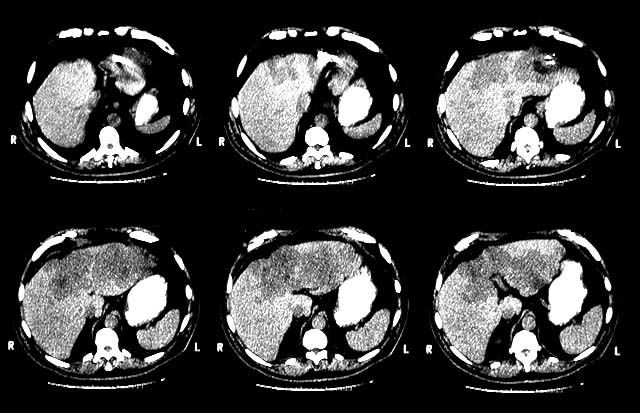
HISTORY: LIVER MASS.
Enhanced axial images were obtained through the abdomen and pelvis in a biphasic fashion following the administration of oral contrast and 150 cc of IV Conray. No old films are available for comparison.
This lesion measures 23.0 cm x 11.0 cm in axial dimension . There is some effacement of the more distal left portal vein, but there is no definite evidence of tumor thrombus. In addition, there is a smaller heterogeneous right lobe liver lesion spanning segments 5 and 6, which measures 8.0 cm x 5.5 cm in axial dimension.
There is no evidence of vascular invasion associated with this separate tumor. The liver is otherwise unremarkable. There is no lymphadenopathy. No other significant abnormalities are identified within the abdomen or pelvis.