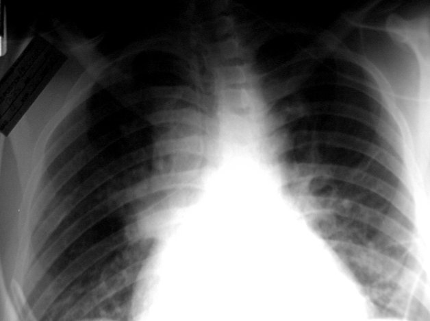
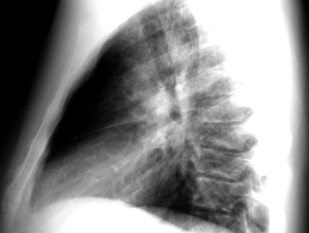
RADIOLOGICAL FINDINGS:
A chest x-ray obtained at the time of admission revealed a right hilar mass (Image 01 and 02), and a CT scan of the chest demonstrated a 4 cm irregular tumor in the right hilum and possible enlargement of mediastinal lymph nodes. A CT scan of the abdomen was negative. Venous Doppler ultrasound was performed to evaluate the bilateral lower extremity edema and revealed no evidence of deep venous thrombosis.
ANTIBIOTIC SUSCEPTIBILITY TESTING