

MICROSCOPIC FINDINGS:
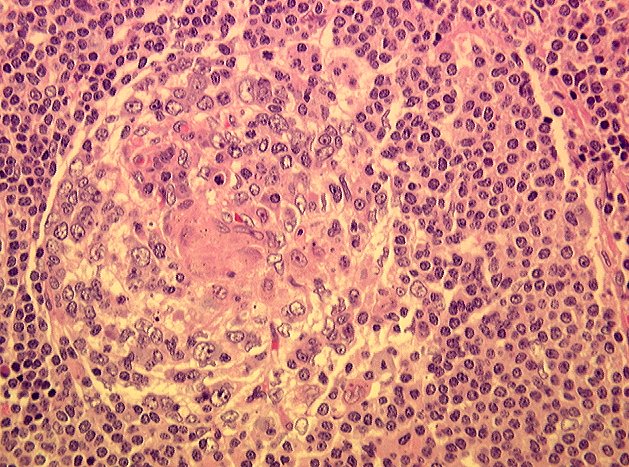
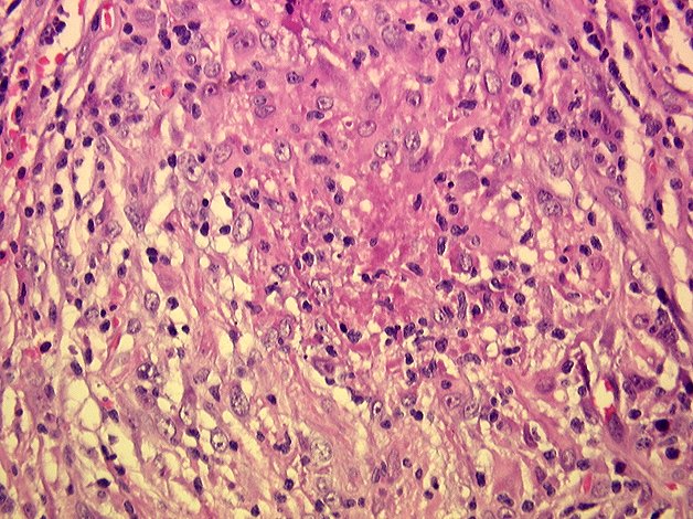
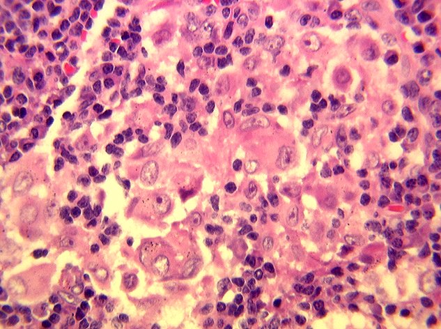
Histological sections of lymph nodes revealed abnormal nodal architecture with focal areas of necrosis (Image 03). Other foci demonstrated multiple granulomata, some of which displayed central caseous-type necrosis. (Image 04, 05, 06, and 07). No carcinoma was seen. Histochemical stains (Acid-fast, Grocott, and Gram) for mycobacteria, fungi and bacteria were negative. Flow cytometry was performed and revealed a polyclonal population of lymphoid cells consistent with a benign or reactive lesion of the lymph node. The preliminary diagnosis of "lymph nodes with necrotizing granulomatous inflammation and eosinophilia" was given.
ANTIBIOTIC SUSCEPTIBILITY TESTING