
![]() Contributed by Hindi Al-Hindi, MD.1, Brian Subach,
MD.2, and Ronald L. Hamilton, MD.1
Contributed by Hindi Al-Hindi, MD.1, Brian Subach,
MD.2, and Ronald L. Hamilton, MD.1
![]() Published on line in February 1997
Published on line in February 1997
This seven year old, right handed, white female presented with a 6-month history of partial complex seizures of increasing frequency, which were eventually controlled with Tegretol. The typical seizure episode was short, lasting only 20-30 seconds, but was followed by a prolonged postictal state. Physical examination was essentially normal and there were no focal neurological deficits. An MRI scan was obtained.
NEUROIMAGING:
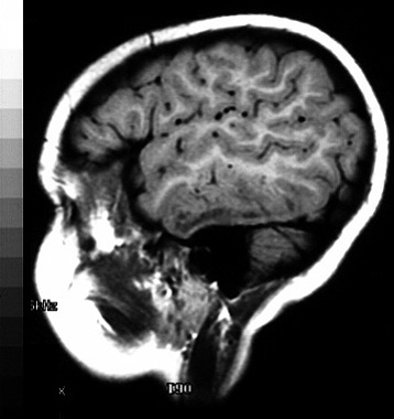
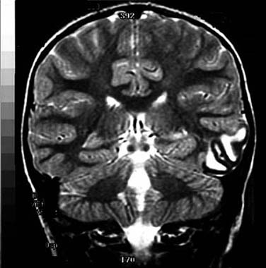
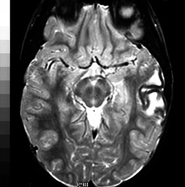
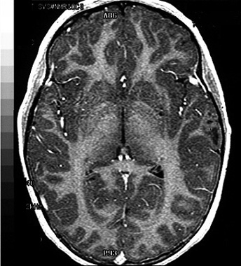
T-1 weighted MRI images demonstrated hypointense white matter signal in the left temporal lobe. The overlying cortex was distinct, but thinned and slightly hyperintense when compared to nearby cortical grey matter. T-2 weighted images showed this area to be hyperintense and the lesion was quite distinct on coronal and horizontal cuts. The lesion showed no contrast enhancement