GROSS DESCRIPTION:
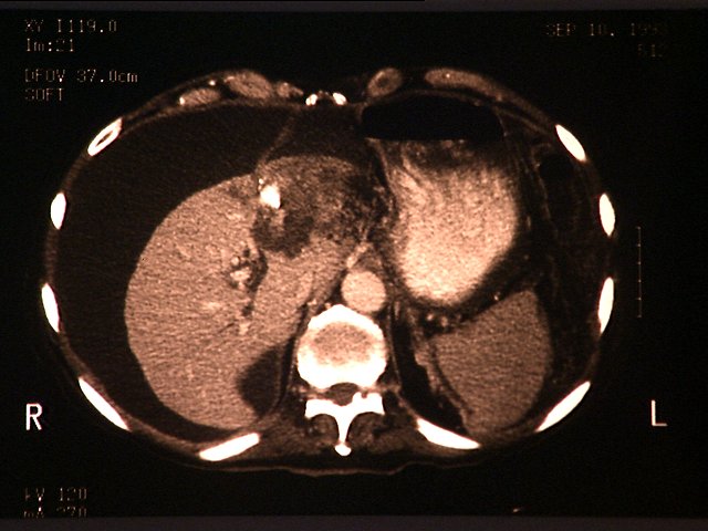
Image 1 is representative of the patient's initial CT scan received on admission to the hospital and demonstrates a 5.0 cm hypodense, calcified mass in the left lobe of the liver. There is a considerable amount of ascites shown on the scan by the distance from the chest wall to the edge of the liver.
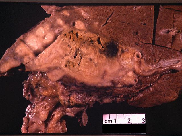
Image 2 is a formalin-fixed section of the liver through the hilus. There is a 5.0 cm partially calcified, necrotic, bile-stained mass involving the hilus and the left lobe. There is also portal vein thrombosis.
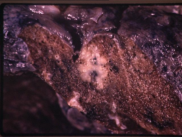
In the lung there are a few firm white nodules present in the parenchyma, from 0.1 to 0.5 cm, a representation of which is presented in image 3.
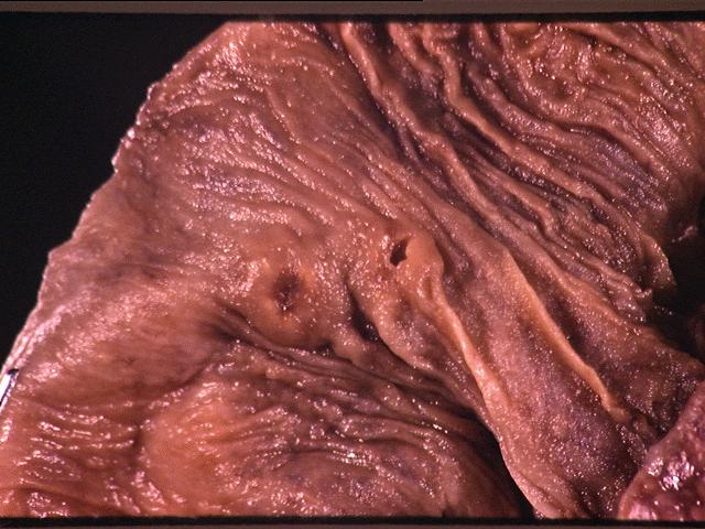
Image 4 shows mucosal ulcerations present in the terminal ileum.