
GROSS DESCRIPTION:
The specimen is received unfixed in two parts.
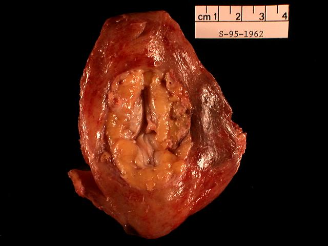
Part 1:
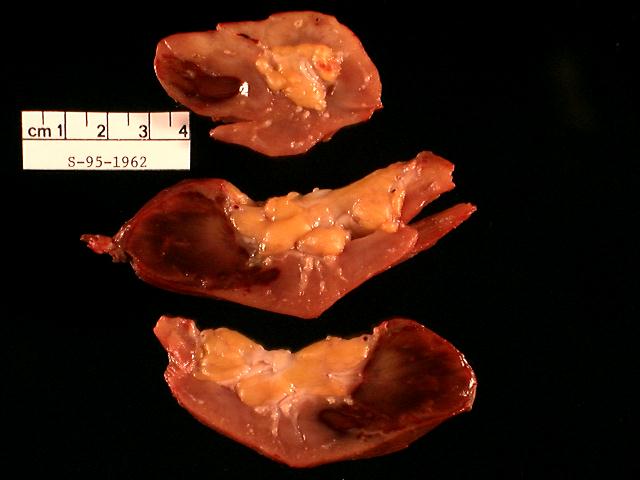
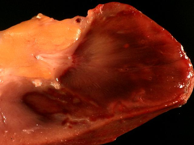
Part 1 is labeled "right allograft kidney" and consists of a 110 gram allograft nephrectomy, 9.0 x 7.0 x 3.0 cm. The cortical surface is irregular and pink-gray with a deeply hemorrhagic 7.0 x 3.0 cm. area at the anterior aspect. Much of the hilum has been previously excavated. The arterial branches are gray-yellow with some narrowing noted. The pelvis is pink-gray, slightly edematous and grossly free of obstruction. The surrounding peripelvic fat is pink-yellow and otherwise unremarkable. The cut surface of the specimen consists of pink-gray and hemorrhagic, atrophic cortex and medulla at the posterior aspect. A deeply hemorrhagic 7.0 x 3.0 x 2.5 cm. comprises shrunken renal cortex and underlying medulla. Severe occlusion of the arterial tributaries is noted within the hemorrhagic parenchyma and also at the periphery. The cortex ranges from 0.5 cm. in thickness at the area of infarction to 1.1 cm. at the convexity. The grossly viable areas of medulla are pink-gray and striated. Several cysts less than 0.1 cm. in greatest dimension are noted in the superior and anterior poles. The pelvis is pink-gray and focally hemorrhagic. The peripelvic fat at the superior pole has a 1.0 x 1.0 cm. area of hemorrhagic, infarction. Adjacent to the infarct an occluded renal artery is noted. Gross photographs are taken. Representative tissue is frozen in bulk and OCT. Additional tissue is submitted in Karnofsky's fixative for possible future electron microscopic evaluation. Sections are submitted.
Part 2:
Part 2 is labeled "subcutaneous lymph node right abdomen" and consists of a 1.5 x 1.0 x 0.8 cm. of pink-gray, fibrotic, encapsulated lymph node with a smooth, pink-gray, fibrotic, cut surface. One half of the lymph node is frozen in OCT, the remainder entirely submitted labeled 2A.