MICROSCOPIC DESCRIPTION:
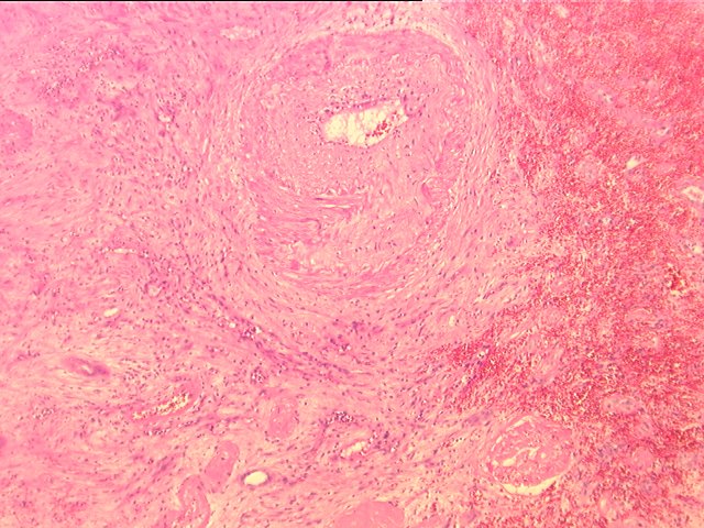
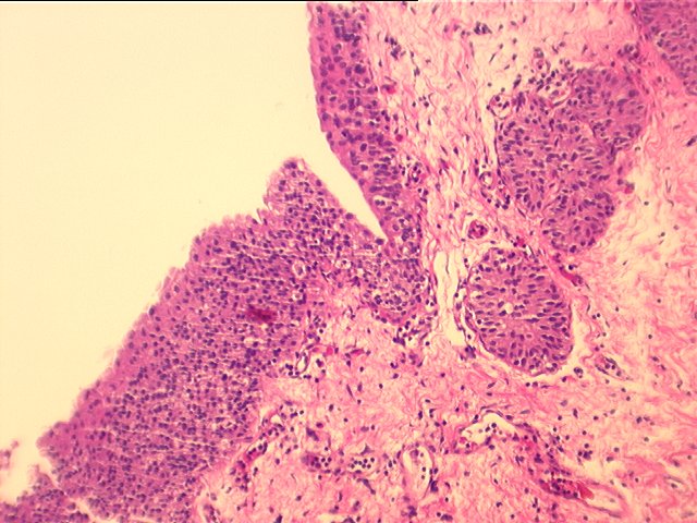
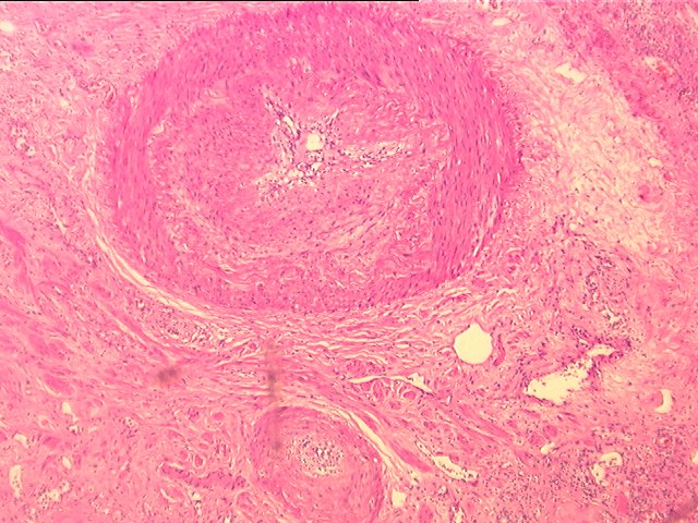
The kidney shows areas of prominent interstitial fibrosis with mild to focal moderate lymphocytic infiltration and alternating hemorrhagic and infarcted areas. Occasional eosinophils are seen. Focal tubulitis is seen, but otherwise the tubules are dilated and demonstrate epithelial reactive changes, focal calcification and in areas thyroidization and transitional epithelial metaplasia. Most of the glomeruli are totally sclerotic with the remainig demonstarting mesangial expansion and capillary loop thickening. The infarcted areas display ghost cells and cellular debris. The most striking changes are seen in the arteries. In all the levels there are variable degrees of obliterative arteriopathy, represented by fibrointimal hyperplasia and foam cell deposition. Some of the arterial branches contain also a mild lymphoplasmacytic infiltrate in the intima, enriched with occasional eosinophils. Section 1B, from the pelvis of the hilum, shows mild focal hyperplasia of the transitional epithelium and mild chronic inflammation. Section 1A, from the perihilar vessels the arterial branches, show moderate fibrointimal hyperplasia. Overall, the changes are suggestive of a chronic rejection process with superimposed component of cellular vascular rejection. No viral inclusions are appreciated. Part 2 consists of a well demarcated round mass composed of mature adipocytes. Mild hemorrhage is seen. No evidence of malignancy is appreciated.