
MICROSCOPIC DESCRIPTION:
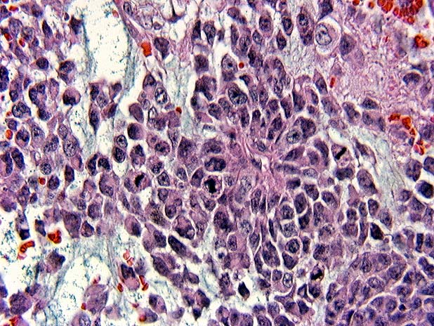
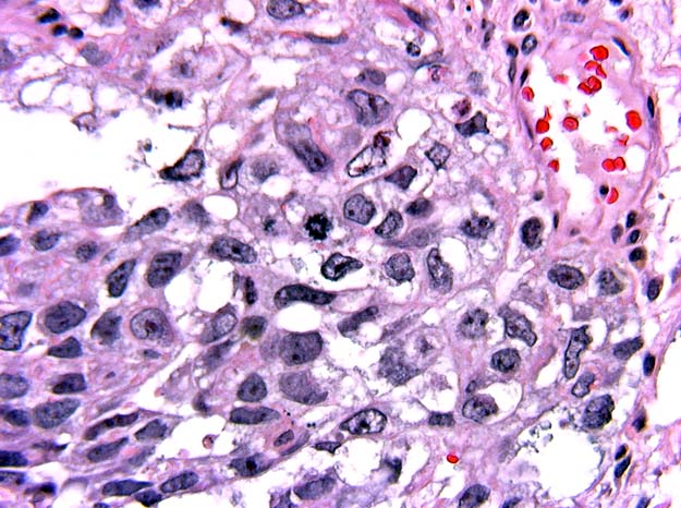
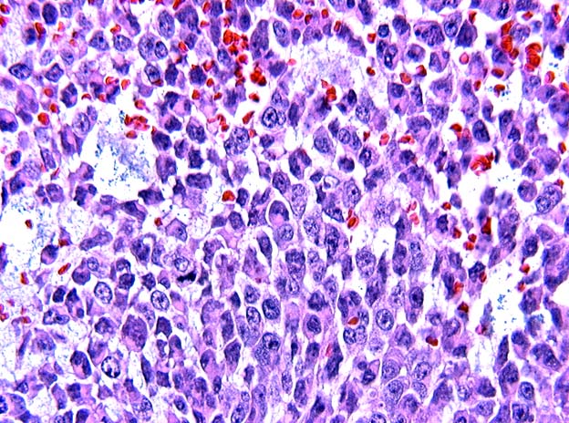
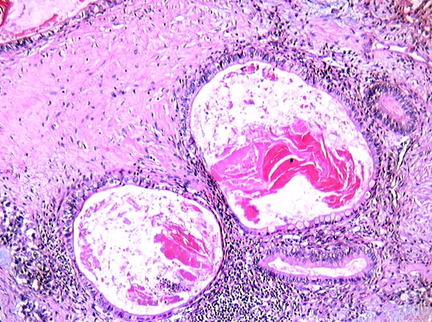
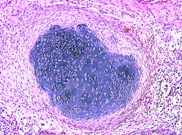
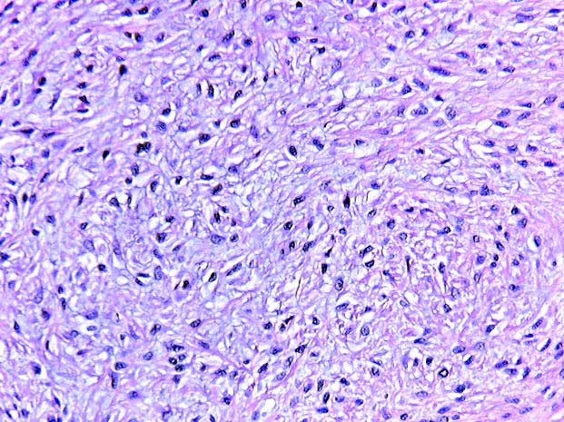
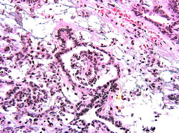
Multiple sections of the tumor reveal a wide variety of histologic patterns. Large areas consist of large, pleomorphic cells with vesicular nuclei and marginated chromatin. Both glandular and solid patterns contain cells with prominent red nucleoli (Images 04, 05 and 06). Other sections display eosinophilic hyaline globules and papillary glomeruloid structures with vascular cores (Schiller-Duvall bodies) (Image 07). In addition, there are teratomatous components with mature and immature cartilage, squamous and glandular areas, mesenchymal stroma, and mature neural tissue (Images 08, 09 and 10). Large areas of hemorrhagic necrosis and islands of syncytial as well as cytotrophoblasts are noted. Angiolymphatic invasion is seen. Invasion of the epididymis and tunica albuguinea is confirmed.
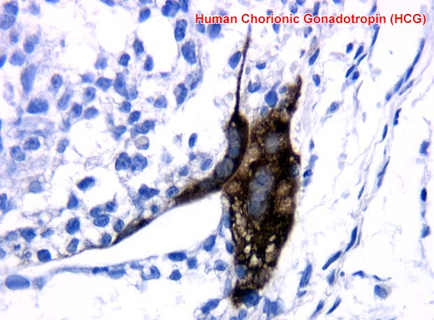
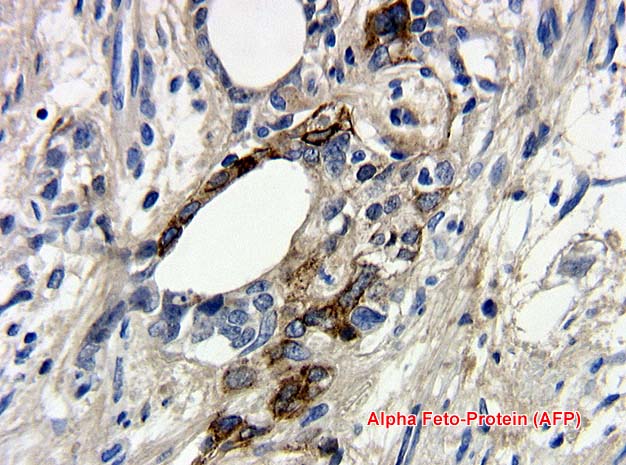
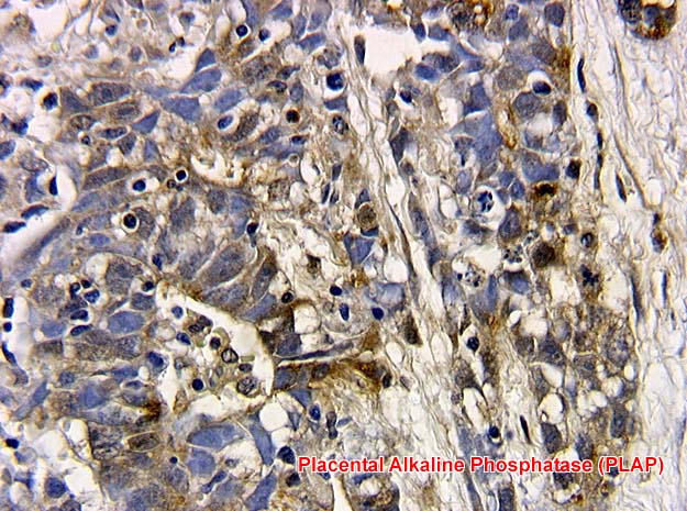
Immunostains reveal the tumor to be focally positive for alpha feto-protein (AFP) (Image 11), human chorionic gonadotropin (HCG) (Image 12), placental alkaline phosphatase (PLAP) (Image 13), and glial fibrillary acidic protein (GFAP) (Image 14).