


MICROSSCOPIC DESCRIPTION:
(6 H&E, 4 MST, 4 PAS, 1 L26 (CD20), 1 CD3, 1 CD43, 1 CD5, 1 CD10, 1 CD68, 1 kappa, 1 lambda slides reviewed.)
The tissue examined by light microscopy consists of renal cortex, renal medulla (Image 01) and an attached portion of renal capsule and perirenal soft tissue.
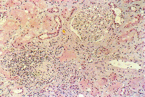
The profiles of up to 27 glomeruli are identified in the paraffin and frozen sections, of which 15 (56%) are globally sclerotic. The remaining glomeruli are normocellular and show no specific light microscopic abnormalities. Silver stains show no evidence of spikes or tram tracking.
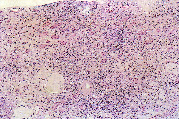
There is moderate, patchy tubular atrophy. Occasional tubules contain granular casts. The interstitium is expanded by a patchy, focally dense, lymphocytic (Images 02, 03) infiltrate (CD3 positive, CD43 positive [Images 04, 05], CD5 positive, CD10 negative, CD20 negative) and moderate interstitial fibrosis. The intrarenal arteries and arterioles show mild chronic changes. There is no evidence of active vasculitis.
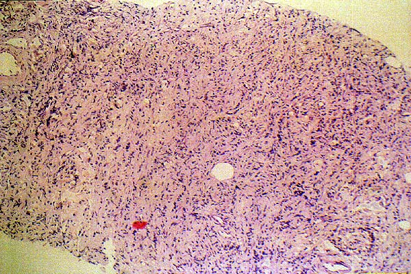
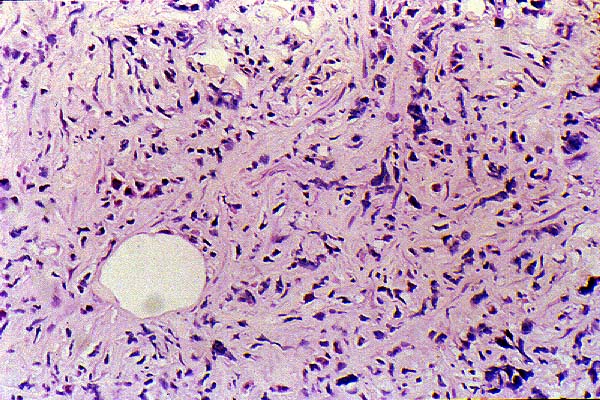
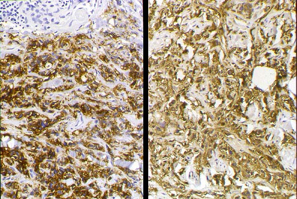
The capsule and attached perirenal adipose tissue are infiltrated by an atypical lymphoid infiltrate (Images 06, 07) containing small and large B-cell lymphoid cells of follicular center origin (CD20 positive, CD10 positive (Image 08), CD3 negative, CD5 negative, CD43 negative), consistent with a B cell lymphoma.
IMMUNOFLUORESCENCE:
The renal tissue examined by immunofluorescence contains six non-sclerotic glomeruli, two sclerotic glomeruli, two small arteries and nine arterioles. Direct immunofluorescence is performed using a panel of ten antisera.