
MICROSCOPIC DESCRIPTION:
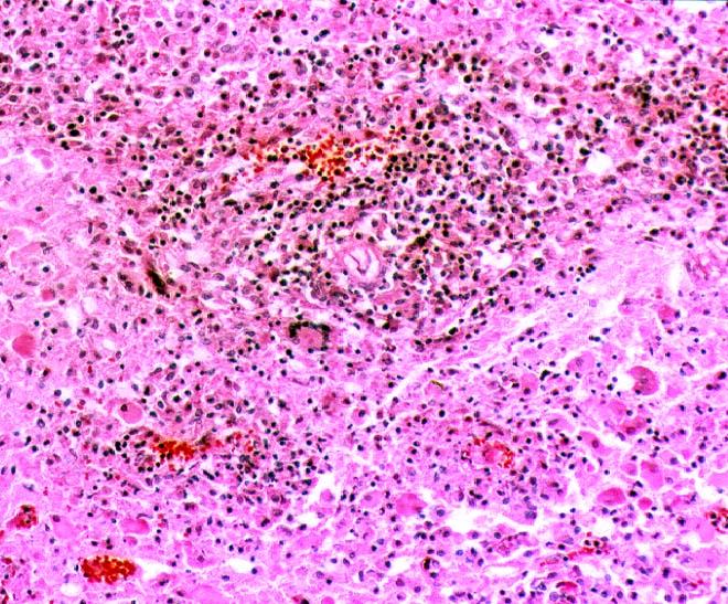
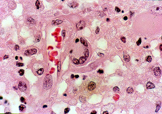
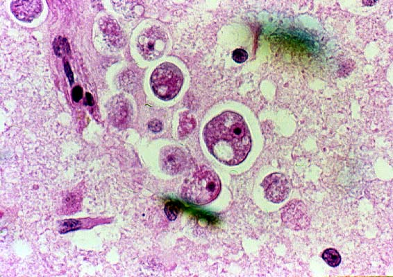
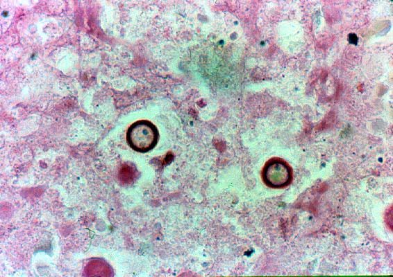
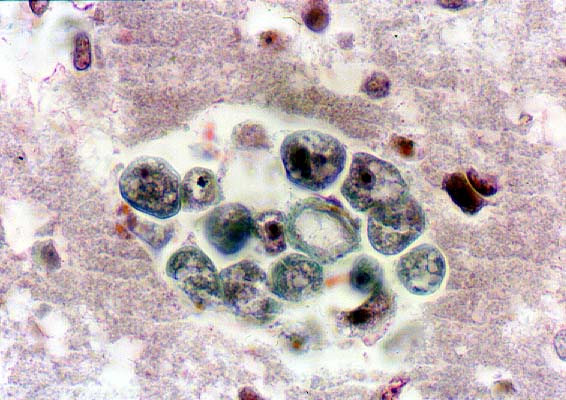
Microscopic examination showed a widespread encephalitis (Image 05), angiitis with focal fibrinoid necrosis, and choroid plexitis. Within and surrounding the foci of necrosis, there were moderate numbers of lymphocytes, a few plasma cells and mononuclear phagocytes, rare neutrophils and scattered petechial hemorrhages. Rare multinucleated giant cells were seen (Image 06). In the areas of encephalitis, there were large cells infiltrating blood vessel walls and perivascular spaces (Image 07). Most of the cells were 8-12 microns in cross section but some were up to 20 microns in diameter. Some of the cells had an angulated or irregular cell outline. They had abundant cytoplasm with numerous, variably sized vacuoles and small nuclei with a dense round basophilic structure surrounded by a halo (Image 08). There were also 10-15 micron rounded structures with a dense, wrinkled, double-contour wall (Image 09). Some of the cells were in aggregates and they had a green hue on Masson trichrome stain (Image 10).
SPECIAL STUDIES:

Electron microscopy demonstrated large, irregularly shaped cells (Image 11) with nuclei showing dense nucleoli, surrounded by finely granular chromatin and well-defined nuclear membranes. The cytoplasm contained numerous mitochondria, lysosomes, vacuoles with myelin figures and prominent Golgi complexes.