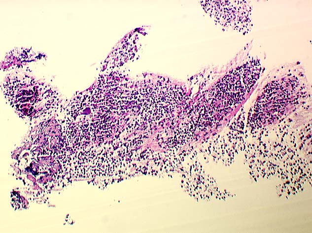


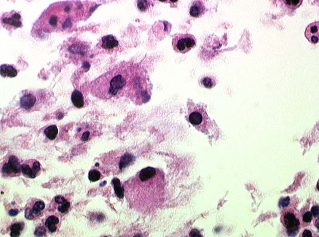


MICROSCOPIC DESCRIPTION:
A single fragment of cartilaginous bronchial wall, multiple fragments of tissue containing lysed red blood cells, and multiple areas containing a fibrinopurulent inflammatory exudate (Images 05 and 06) were identified. Amongst the fibrinopurulent inflammatory exudate, a background of histiocytes containing granular eosinophillic foamy cytoplasm was also present (Images 07 and 08). Whereas acid fast and Grocot stains were negative for microorganisms, a tissue gram stain revealed multiple gram-positive coccobacillary organisms associated with the histiocyte cytoplasm (Images 09, 10 and 11). A diastase-treated PAS stain showed abundant collections of positive staining material within the cytoplasm of the histiocytes (Image 12).