


MICROSCOPIC DESCRIPTION
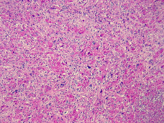
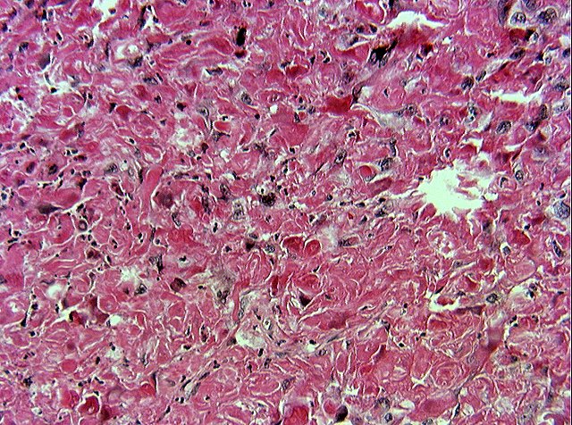
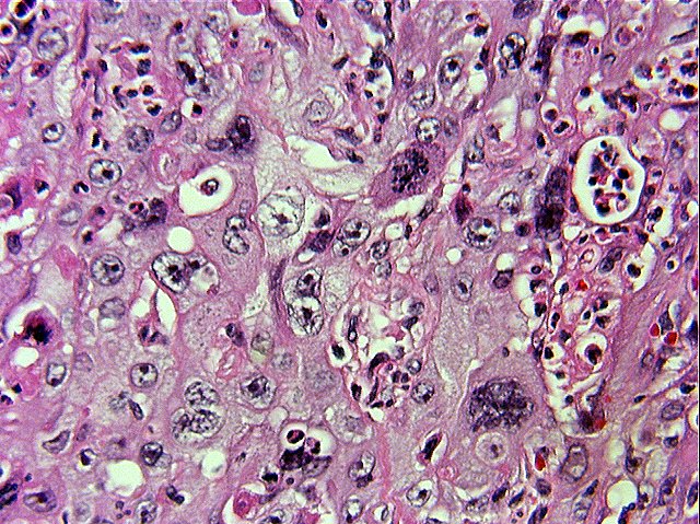
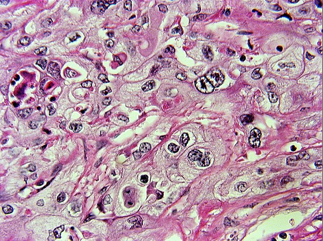
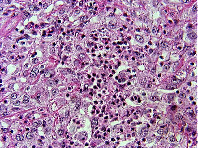
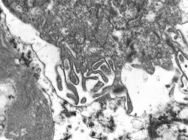
This poorly differentiated carcinoma is characterized by solid sheets of large polygonal tumor cells with eosinophilic ground glass cytoplasm and distinct cell borders. (Image 02 & Image 03) The nuclei are irregular with prominent eosinophilic nucleoli. (Image 03) Mitotic activity is brisk with bizarre tumor giant cells.(Image 04) Focal areas show clear cell differentiation.(Image 05) Fibrous septa are present associated with a lymphoplasmacytic infiltrate with occasional eosinophils. (Image 06) There is a scattered neutrophilic infiltrate associated with the tumor.(Image 07) Focal abortive keratin production is noted as well as focal mucin positivity.(Image 08) Ultrastructurally, there are well developed desmosomes and interdigitating microvilli. (Image 09 & Image 10)