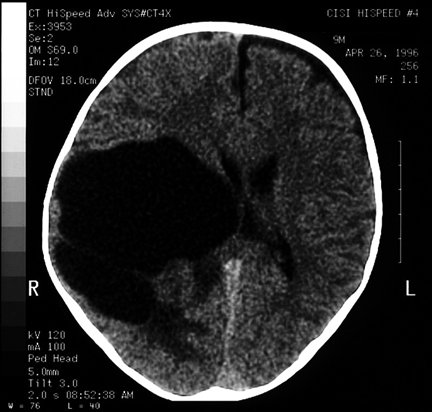

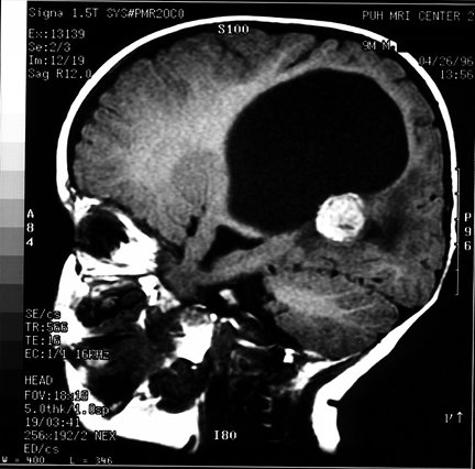


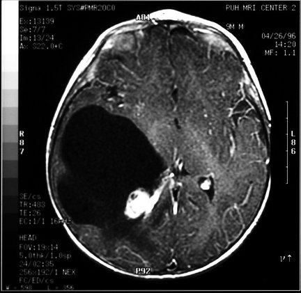
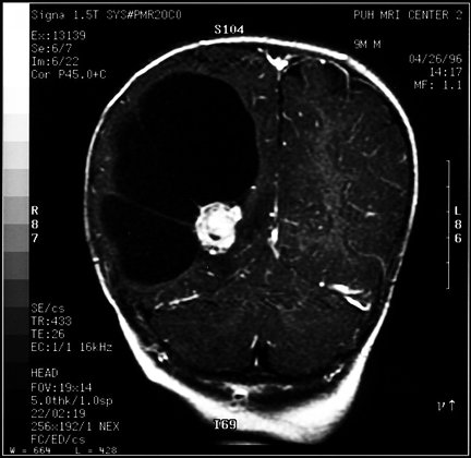
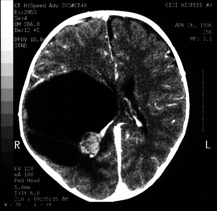
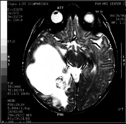
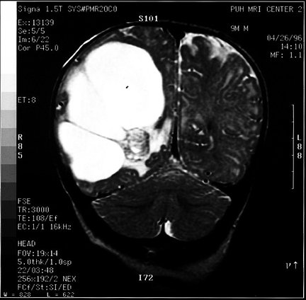
CT scans revealed a huge cystic space in the right hemisphere with a nodule. It was uncertain if this was a parenchymal nodule with surrounding cyst or intraventricular lesion. There was no evidence of calcification. The lesion enhanced brightly with contrast. A sagittal T-1 weighted MRI demonstrates a hyperintense, well circumscribed nodule. On T-2 weighted images the nodule has many hypointense areas on both axial and coronal views. There was strong contrast enhancement of the lesion as seen in these post-contrast axial and coronal studies.
At surgery, a fairly-well circumscribed, heavily pigmented nodule at the trigone was found. It protruded into the ventricle and was surrounded on three sides by choroid plexus. It was completely resected. The neurosurgeon's impression was of a pigmented tumor which might be a progonoma (retinal anlage tumor, or melanotic neuroectodermal tumor of infancy).