RADIOLOGY
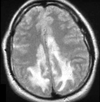
Axial T2-weighted magnetic resonance image just above the lateral ventricles demonstrated multifocal regions of high signal intensity within the white matter, particularly in the parietal and occipital lobes. This abnormality was unassociated with mass affect.