MICROSCOPIC DESCRIPTION
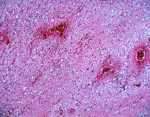
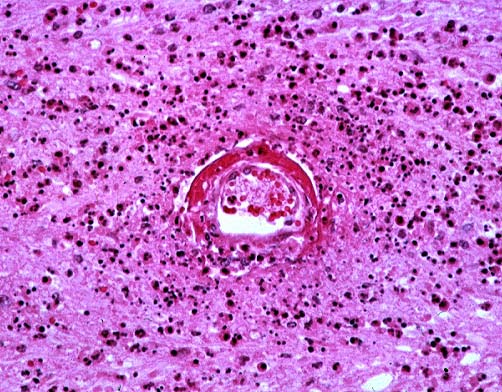
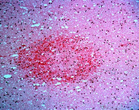
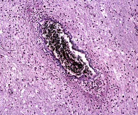
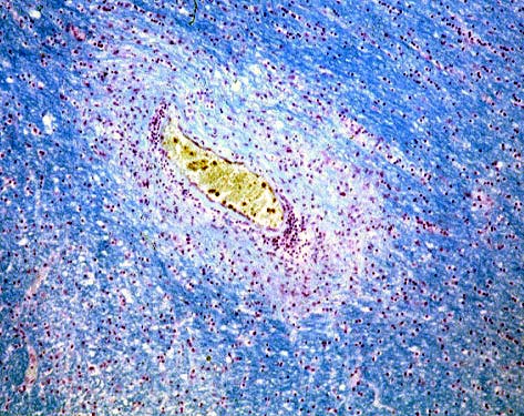
Post-mortem examination revealed a diffusely swollen brain with patches of gray discoloration and scattered petechial hemorrhages within the white matter. Low-power microscopic examination disclosed an inflammatory infiltrate within the centrum ovale with associated vascular necrosis (Image 01). The predominantly neutrophilic infiltrate was focally brisk, and the vasculitic lesions were accompanied by fibrin deposition (Image 02) in the Virchow-Robin space. Scattered ring-shaped hemorrhages (Image 03) were present. Perivascular to confluent foci of demyelination were highlighted by luxol fast blue stain and characterized by the relative preservation of axons (Image 04 and Image 05).