Case 978-- A 37-Year-Old Woman with Progressive Right Side Ptosis for One Month

 Contributed by Hui-Yuan Su, MD 1,2, Chun-Chieh Wu, MD3, Aij-Lie Kwan, MD,PhD1,2,4,5, Chih-Lung Lin, MD,PhD2,4,5, Ann-Shung Lieu, MD,PhD 2,4,5, Yu-Feng Su, MD2,6
1Department of Surgery, Kaohsiung Medical University Hospital, Kaohsiung Medical University, Kaohsiung, Taiwan
2Department of Neurosurgery, Kaohsiung Medical University Hospital, Kaohsiung Medical University, Kaohsiung, Taiwan
3Department of Pathology, Kaohsiung Medical University Hospital, Kaohsiung Medical University, Kaohsiung, Taiwan
4School of Medicine, College of Medicine, Kaohsiung Medical University, Kaohsiung, Taiwan
5Graduate Institute of Medicine, College of Medicine, Kaohsiung Medical University, Kaohsiung, Taiwan
6Graduate Institute of Clinical Medicine, Kaohsiung Medical University, Kaohsiung, Taiwan
CLINICAL HISTORY


A 37-year-old woman had bilateral blurred vision with hazy vision gradually from periphery to center for two months. There was mixed horizontal and vertical diplopia. Ophthalmologic treatment did not help. We she came to us, an Humphrey visual field test disclosed bilateral temporal hemianopia. CT showed hyperdensity and heterogeneously enhanced sellar lesion with size 2.57 x 1.96 x 3.63 cm and supra-sellar extension. MRI T1 showed sella tumor with isointense signal mixed with the hyperintense part (Fig 1a). MRI T2 scans showed compression of the optic chiasm. With contrast, there was heterogeneous enhancement in the tumor. The clivus showed a heterogenously enhancing lesion was noted on T1 with contrast (Fig 1b).
She underwent endoscopic endonasal tumor resection and the pathology showed a pituitary adenoma. Post-Operative CT showed residual tumor at the sellar turcica (Fig 1c). There was central diabetic insipidus which improved after desmopressin treatment. She was then discharged. Unfortunately, one month later, she suffered from progressive diplopia, right side ptosis, impairment of right side visual acuity, with dizziness, and headache. Follow-up clinic visit revealed palsies in the right side optic nerve, oculomotor nerve, trochlear nerve, and abducens nerve. Serial brain CT showed rapid progression of the sellar tumor with suprasellar extension and compression on the midbrain at 1 and 2 months after her initial operation (Figs 1d and 1e respectively). MRI T1 with enhancement disclosed strongly enhanced sellar tumor (Fig 1f). Laboratory data disclosed normal range of βhCG
After discussion with patient and family, further surgical intervention was suggested. Operation of pterional approach and subtotal removal of the tumor was performed. Intra-operatively, soft, yellowish and easy bleeding tumor was noted. After the operation, appropriate recovery condition with clear consciousness was noted for three weeks. After that, deterioration of the consciousness, irregular respiratory pattern and bilateral dilatation of the pupils without light reflex were noted since the 18th post-operation day. After the second operation CT scan showed rapid regrowth of the tumor with suprasellar extension and brainstem compression (Fig 1g). On the 24th post-operation day, we informed family the definite pathology diagnosis. After discussion with family, they decided to arrange supportive care to this patient. Then, this patient was expired on the 32th post operation day.
MICROSCOPIC PATHOLOGY
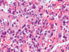
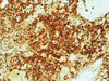
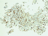
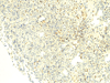
Histopathological examination revealed tumor cells with large, vesicular, rounded to bean-shaped nuclei, prominent nucleoli, and abundant cytoplasm, arranged in sheets or in a solid trabecular pattern (Fig 1h). The tumor cells are positive for EMA (Fig 1i), and the Ki-67 labeling index is around 60% (Fig 1j). In addition, INI-1 loss is also noted (Fig 1k). What is your diagnosis?
FINAL DIAGNOSIS


)
)
)
)
)
)
)
)
)
)
)