Case 951-- A 31-Year-Old Man with Slowly Progressive Limb Weakness and Respirtory Insufficiency

 Contributed by Akinori Uruha, MD, PhD1, 2, Yukiko K. Hayashi, MD, PhD1, 2, 3, Madoka Mori-Yoshimura, MD, PhD4
Yasushi Oya, MD4, Masahiro Kanai, MD4, Miho Murata, MD, PhD4, Ichizo Nishino, MD, PhD1, 2
1Department of Genome Medicine Development, Medical Genome Center, and
2Department of Neuromuscular Research, National Institute of Neuroscience, National Center of Neurology and Psychiatry (NCNP), Japan
3Department of Pathophysiology, Tokyo Medical University, Japan
4Department of Neurology, National Center Hospital, NCNP, Japan
CLINICAL HISTORY
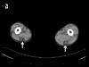
A 31-year-old man, with no family history of neuromuscular diseases, received muscle biopsy for slowly progressive muscle weakness. The first symptom he had was difficulty dorsiflexing his feet at age 20 years, which was followed by gradual development of gait disturbance. At age 27 years, he started using a handrail to climb up stairs. At age 28 years, he developed dysphagia for liquid, in addition to difficulty raising arms, which led him to have aspiration pneumonia later in the same year. At age approximately 30 years, he developed dyspnea on exertion. Arterial blood gas analysis revealed hypoxemia (65 mmHg, at room air) and hypercapnia (82 mmHg), with a vital capacity decreased to 780 ml, leading to the diagnosis of chronic type 2 respiratory failure, and non-invasive ventilation was started. At age 31 years, he was unable to walk without aid and required a wheelchair for long distances. Physical examination revealed moderate muscle weakness and atrophy in an asymmetric limb-girdle distribution, together with marked muscle atrophy in the tibialis anterior muscles and weakness in ankle dorsiflexion with Medical Research Council grade 1. Mild neck muscle weakness was also noted. Serum creatine kinase level was 375 IU/L (normal: <287 IU/L). Electromyography showed myopathic changes. Skeletal muscle CT demonstrated remarkable fat tissue replacement in the semitendinosus muscles (figure 1a, arrows) at the thigh level.
PATHOLOGY
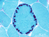
Skeletal muscle pathology from the biceps brachii demonstrated myopathic changes with moderate fiber size variation, mildly disorganized myofibrillar networks, some fibers with cytoplasmic bodies, which were often located in line in subsarcolemmal region of a muscle fiber (figure 1b, modified Gomori trichrome stain), and a few fibers with rimmed vacuoles. What is your diagnosis? What is the next test to confirm it?
FINAL DIAGNOSIS


)
)