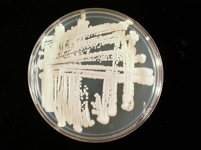
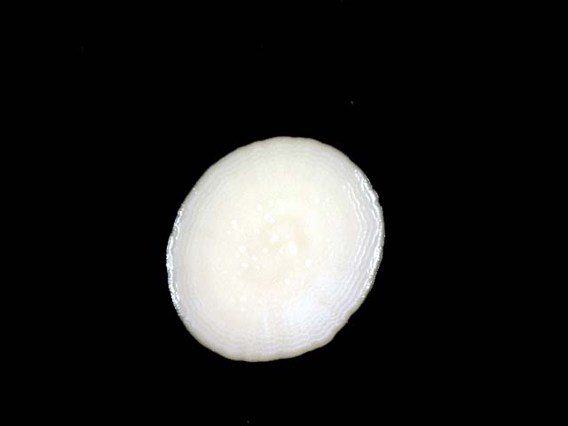

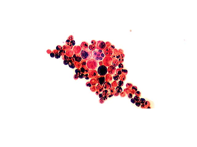
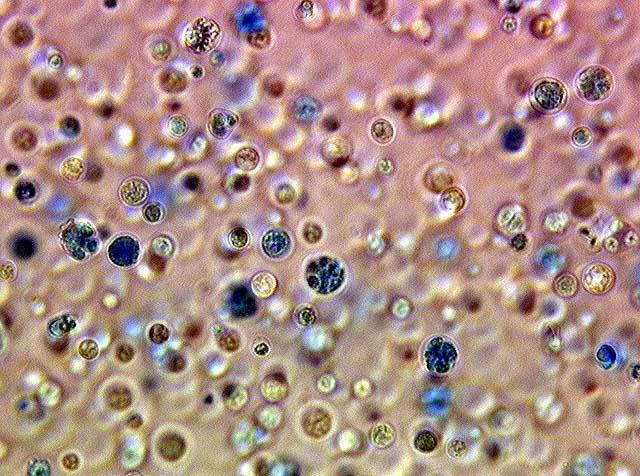
Prototheca grows easily on glucose containing media and is typically grown on sabouraud's agar, but it will not grow on media that contain cycloheximide. The colonies (Images 04, Image 05) appear yeast-like and are smooth, moist, and white to cream in color. The algae grows best at 30 C and growth is inhibited at 37 C (1,2). Prototheca are unicellular organisms that are typically round to oval and 8-16 um in diameter, though they can range from 3 to 30 um depending on the species and the degree of maturation. The organisms reproduce by a mother cell undergoing internal cleavage. This results in the internal accumulation of smaller endospores surrounded by the wall (or theca) of the mother cell. The mother cell then bursts, releasing the endospores and the cycle is repeated. The endospores are 4 to 5 um in diameter and can number from two to 50 (2). These endospores give Prototheca its characteristic "cart wheel" appearance of a round structure with internal septations. The larger forms can have very thick walls. The algae are basophilic and have been reported to be Gram positive (2), though in this case the theca are Gram negative and the endospores Gram positive (Image 06, Image 07). PAS staining highlights the starch granules which are occasionally present.
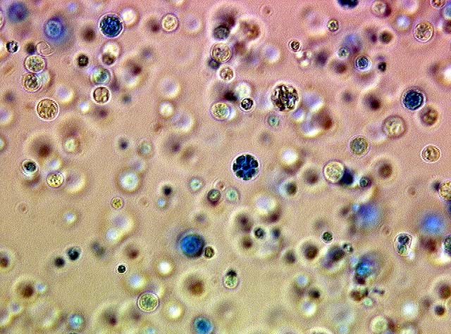
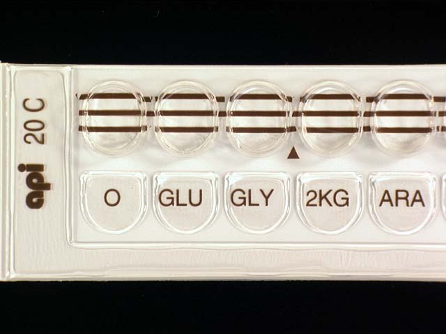
Prototheca can be identified by its colony morphology in conjunction with its characteristic microscopic appearance when examined via wet mount or a lactol-phenol cotton blue preparation (image 08, Image 09). Speciation, however, requires further analysis and relies on diameter, assimilation of various sugars and the presence or absence of an external capsule (1,3,4). Commercial strips (image 10) are available that can also be useful in species identification. Using an API 20C, this case was speciated to P. wickerhamii (5).