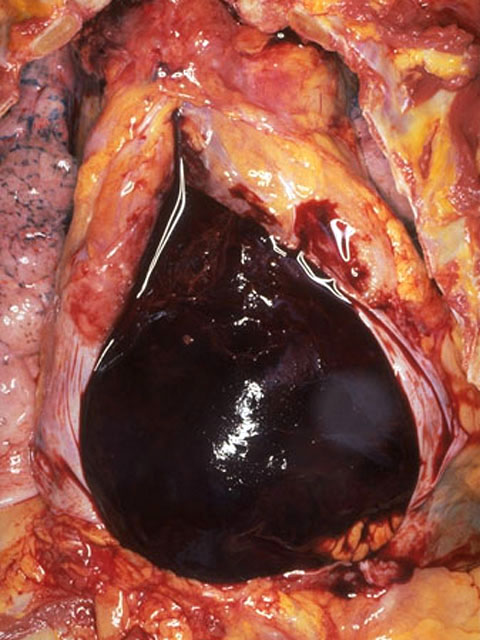
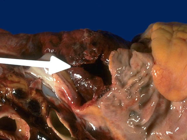
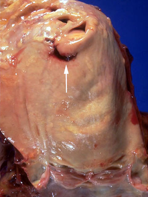
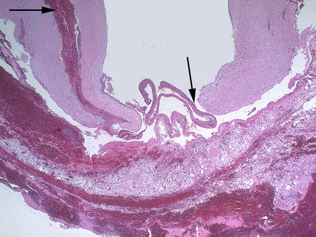
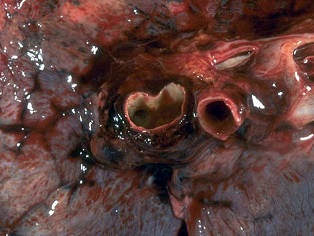

At autopsy, the pericardial cavity was opened to reveal 300 ml of clotted and 330 ml of liquid blood that produced cardiac tamponade. This blood emanated from an area on adventitia of posterior aortic arch. The heart weighed 350 gm and the coronary arteries showed moderated atherosclerosis with no significant lumenal narrowing. Upon opening the heart and aorta, an 1 cm tear was present in the aortic arch just proximal to the great vessels. This tear microscopically revealed dissection through the media, as seen below the center arrow, as well as dissection into the outer media, as seen at the upper left arrow. The aorta demonstrated marked atherosclerosis, including the arch and thoracic portion, as well as abdominal aorta. There was no microscopic evidence of cystic medial necrosis with mucin and elastic tissue stains. Blood had dissected into and through the adventitia of the aorta posteriorly, filling the pericardial cavity. Blood dissected into the mediastinum, widening it, and from there into the pleural cavities, where there was 500 ml on the right and 500 ml on the left of bloody fluid. Blood dissected into the hilum of the right lung and the left lung. Blood dissected along the outer media and adventitia of the aorta to the coronary ostia, compressing them slightly, and to the great vessels, markedly compressing the right carotid artery.