


GFAP
Synaptophysin
Synaptophysin
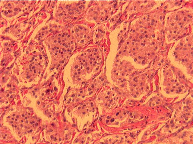
Microscopic examination reveals a tumor composed of homogeneous cells with oval to round nuclei and moderate eosinophilic cytoplasm. The tumor cells show a "zellballen" arrangement, with nests lined by sustentacular cells. GFAP and Synaptophysin stains are positive in the tumor cells.