DIAGNOSIS
Bifocal myxopapillary ependymoma (WHO grade I).
DISCUSSION
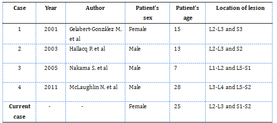
First described by Kernohan in 1932, Myxopapillary ependymoma (MPE) is a variant of ependymoma, occurring almost exclusively in the conus medullaris or ?lum terminale region of the spinal cord, although there are other sites including the cervicothoracic spinal cord and intracranial region. It is de?ned as a well-circumscribed, slow-growing glioma that has a favorable prognosis and long-term survival, which is categorized as WHO grade I. The male-female ratio of this tumor is 2.2:1, with a mean patient age of 36 years (6). They mostly appeared as solitary lesions but not bifocal ones. We recently encountered a relatively rare case of bifocal MPE in a 25-year-old woman, and it is reported here. There are only four other cases of bifocal MPE of the filum terminale after an extensive review of the literature (2,3,7,8) (see table below).
Clinically patients usually may feel low back pain or lumbosciatic neuralgia. Histological features of MPE are very distinct. It is composed of abundant papillary arrangement of well-differentiated cuboidal or columnar tumor cells. These cells surround central cores that contain small blood vessels and/or myxoid stroma, forming perivascular pseudorosettes. Cytological atypia, mitotic activity and necrotic foci are usually absent. The immunohistochemistry for the tumor cells often show the strong and diffuse positivity in GFAP, S-100 and vimentin. Cytokeratin, EMA and NF are not expressed. The Ki-67 (MIB-1) labeling index is always low. The main considerations in the clinical and radiological differential diagnosis generally include chordoma, schwannoma, meningioma paraganglioma, mesothelioma, myxoid chondrosarcoma and papillary adenocarcinoma, which are easily excluded histologically and immunohistochemically.
MPE arises from embryonic rests of ependymal cells (5). Whether these double lesions are bifocal or spreading is controversial. Hallacq et al (3) considered the two lesions proximal and distal on the terminal ?lum might represent one end of a spectrum, the other end being the giant tumor of the cauda equina region. And he confirmed the absence of tumor tissue strand in the terminal filum between both lesions. Nakama et al (8) suspected it is possible that a double lesion is one aspect of the tumor progression. McLaughlin et al (7) proposed patients with double MPE may potentially have an early genetic alteration in ependymal stem cells within filum terminale and the subsequent accumulation of different genetic alterations may explain the occurrence of two different lesions each harboring a different genetic signature. Based on these views, we tend to think that bifocal MPE are independent lesions. The exact mechanism of development of multiple MPEs needs to be further proved.
The recurrence rate for MPE in patients is low and prognosis is excellent if tumor total resection can be achieved. Though after gross total resection, the rate of recurrence of MPE is 10 to 19%. FISH analysis of the status on chromosome 1p may contribute to assessing recurrence risk in MPE (9). Additional radiotherapy was once recommended only in cases of which were recurrent and/or difficult to be resected completely. Recently, Bagley et al (1) demonstrated additional radiotherapy or chemotherapy was no benefit by studying the clinical course of 52 MPE including 38 adult patients and 14 pediatric patients. And his research suggested that PDGFR? may be a potential therapeutic target in recurrent MPE.
Despite MPE is ranked as a low-grade tumor because of its benign characters, a case of intracranial metastasis has been occasionally reported (4). Therefore MPE patients should be followed with craniospinal axis imaging. The present case also requires prolonged follow-up close observation because only resection was performed.
Table: Clinical material summary of bifocal myxopapillary ependymoma of the filum terminale in the literature
REFERENCES
![]() Contributed by Yang Wen-sheng MD, Shao Wei MD, Liu Dan MD, Hu Jun-cheng MD, Lin Zhen MD, Qi Pei-lin MD, Wang Cheng-Feng MD, Ji Tian-hai, MD.
Contributed by Yang Wen-sheng MD, Shao Wei MD, Liu Dan MD, Hu Jun-cheng MD, Lin Zhen MD, Qi Pei-lin MD, Wang Cheng-Feng MD, Ji Tian-hai, MD.