![]() Contributed by Russell Silowash, DO and Bruce Rabin, MD,PhD
Contributed by Russell Silowash, DO and Bruce Rabin, MD,PhD
PATIENT HISTORY
A 66 year old married, Caucasian male with a history of HIV/AIDS presents to the emergency department with a chief complaint of back pain for the past 3 weeks and hemoptysis for the past few days. The pain is sharp, in the left side of his lower back, and does not radiate or cause weakness or numbness. The pain is exacerbated with movement and minimally relieved with pain medication. The patient denies fevers, night sweats, and weight loss. His HIV is managed with Atripla. His HIV RNA levels are undetectable. His family history is significant for his mother having pancreatic cancer, his father having prostate cancer, and his brother acquiring tuberculosis at the age of 14. The patient is a retired maintenance man and lives at home. He has a 15 pack year smoking history. He denies alcohol and intravenous drug use. The physical exam is significant for tachycardia and tenderness over the left rib cage and flank. A straight leg test is positive at 30 degrees, reproducing the patient's flank pain.
IMAGING
A CT scan of the chest, abdomen (Figure 1), and pelvis reveal lytic lesions involving the vertebrae, bilateral iliac wings, and sternum. There is a non-displaced fracture of the left posterior tenth rib. A CT scan of the head reveals lytic lesions of the calvarium.
LABS
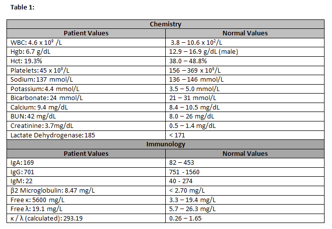
Table 1 shows baseline lab values. The patient is anemic, thrombocytopenic, and has renal insufficiency. His lactate dehydrogenase is also elevated. Immunology studies reveal decreased levels of IgG and IgM as well as increased β2 microglobulin levels and elevated free Kappa (κ) light chain levels. Free Lambda (λ) light chain levels are within the normal range.

Figure 2 shows the serum protein electrophoresis results along with immunofixation. Serum protein electrophoresis shows a definitive band in the gamma region that corresponds with the band in the κ column on immunofixation. There are no bands within the IgG, IgA, or IgM immunofixation columns. These results rule out IgGκ, IgAκ, and IgMκ. IgE and IgD are not routinely tested for in our labs, but when a κ band without an associated heavy chain is seen, IgD and IgE immunofixation is performed to test for IgDκ and IgEκ. Immunofixation results for IgDκ and IgEκ are exhibited in Figure 3. A definitive κ band is present. There are no bands in the IgD or IgE columns, ruling out IgDκ and IgEκ. These results suggest free κ chains in the blood. Urine protein electrophoresis and immunofixation results are shown in Figure 4. Urine protein electrophoresis shows large amounts of protein in the urine, with a majority of proteins lying in the gamma region of the gel. κ bands are present, signifying free κ chains in the urine. Serum free κ and λ levels are quantified, and a κ /λ) ratio is calculated, yielding a value of 293.19 (Normal range: 0.26 - 1.65).
A bone marrow biopsy is performed and reveals reduced trilineage hematopoiesis and hypercellular bone marrow involved by neoplasm. Kappa-restricted plasma cells comprise approximately 90% of cells.