


MICROSCOPIC DESCRIPTION
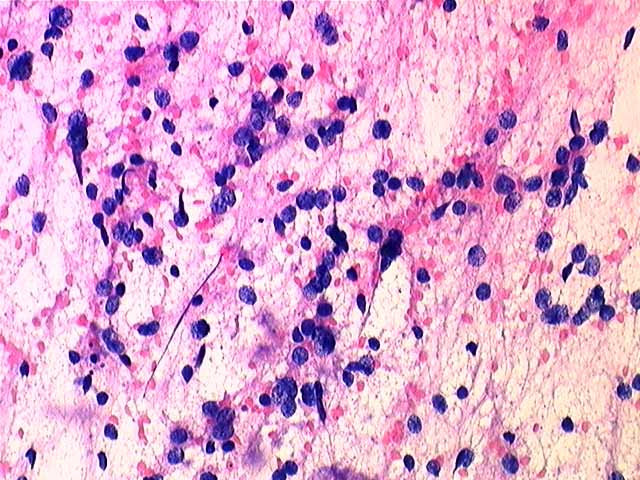
Touch preparation at intraoperative consult showed numerous cells, often appearing individually, but also present in clumps. The cells had minimal cytoplasm and round to slightly oval nuclei with delicate chromatin and minimal pleomorphism. The eosinophilic background was primarily granular in nature, although in some areas it appeared more fibrillar. There were no mitoses or necrosis. The impression was of a primary CNS neoplasm with a differential given of central neurocytoma vs. oligodendroglioma vs. esthesioneuroblastoma.
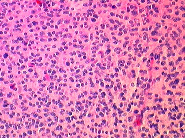
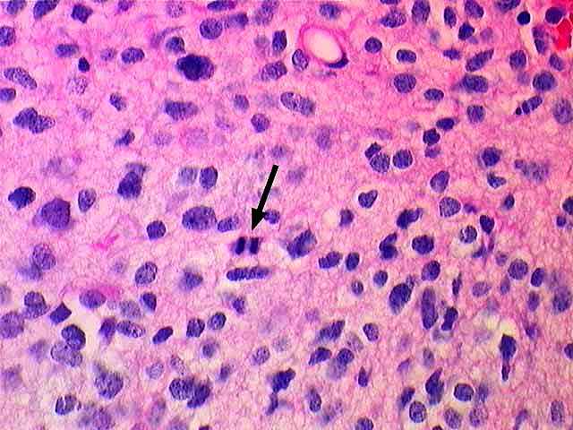
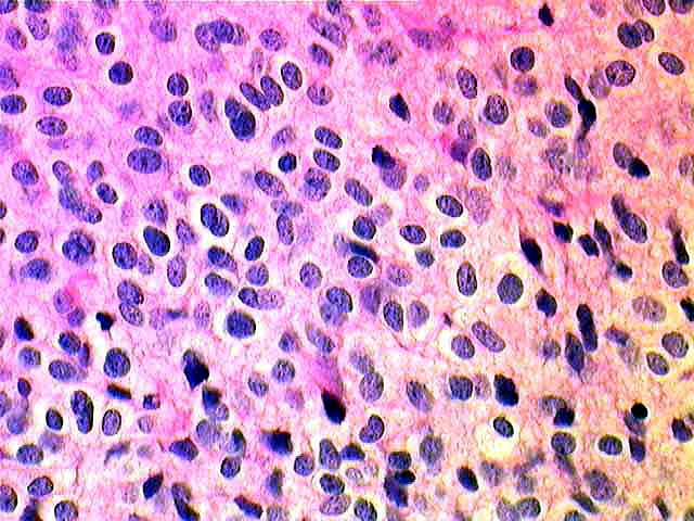
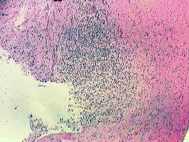
Paraffin embedded material showed a cellular neoplasm containing a fairly monomorphic population of cells with round to slightly oval nuclei containing delicate chromatin. There were no nucleoli and only rare mitotic figures. In a few areas the cells were more pleomorphic and sometimes clumped together, but there were no true ganglion cells and no Homer-Wright rosettes. In many areas there was clearing around the nuclei reminiscent of that seen in oligodendrogliomas. There were rare calcifications. There was no endothelial proliferation or necrosis. The tumor invaded the surrounding brain in a broad front pattern rather than diffusely infiltrative.
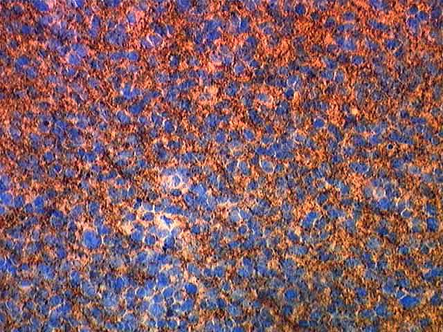
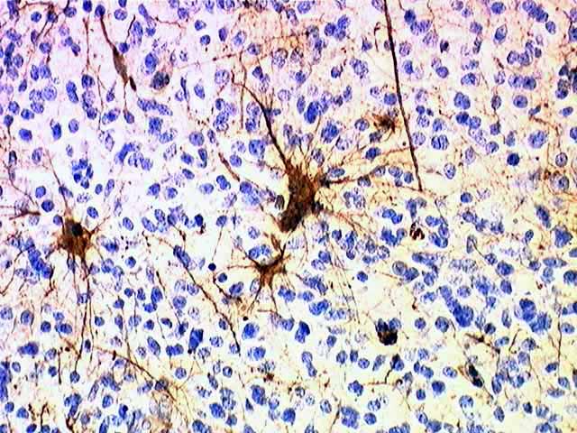
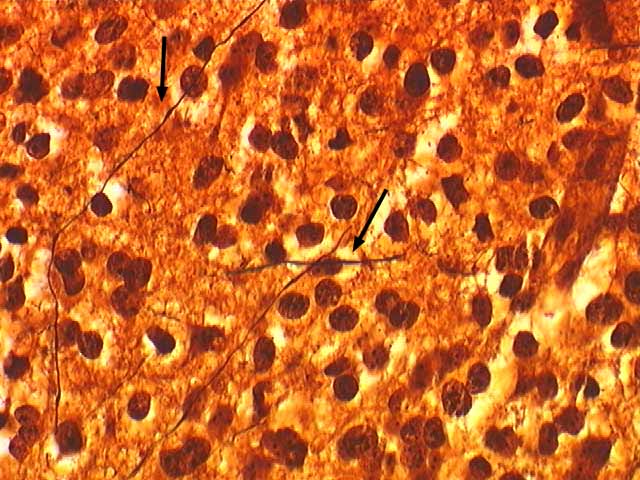
Immunohistochemical stain showed strong, diffuse positive staining for synaptophysin. GFAP stains showed scattered positive cells within the tumor which resembled reactive astrocytes. These same cells stained with S-100. A Bielschowsky stain showed neuronal processes within the tumor.
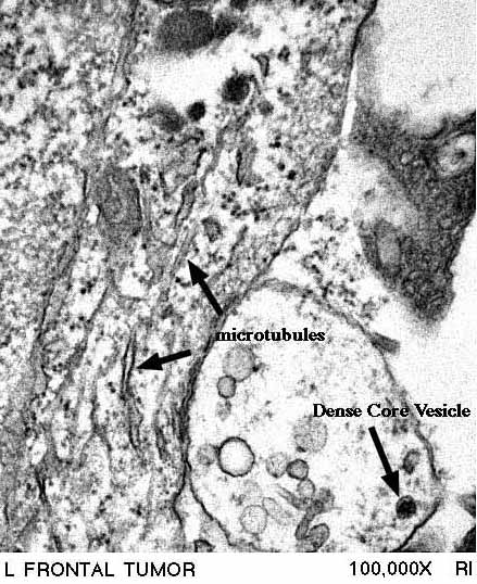
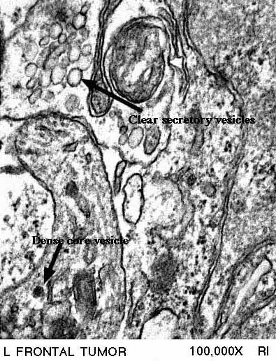
Electron microscopy demonstrated tumor cells to contain dense core granules and microtubules as well as secretory vesicles. No synaptic structures were identified.