(Adapted from Gastrointestinal and liver pathology - Foundations in diagnostic pathology, 2005)
FINAL DIAGNOSIS
Metastatic gastrointestinal stromal tumor (GIST), epithelioid type.
GIST -Disease Fact Sheet
Gastrointestinal stromal tumors (GISTs) arise in the GI tract and occasionally separately within the abdomen. GISTs are mesenchymal tumors displaying differentiation along lines of the interstitial cells of Cajal (ICC), whose function is involved with gut motility. GISTs affect men and women roughly equally, over a wide age range; however, 75% of cases occur in adults aged over 50.
GISTs generally arise as solitary tumors of the stomach (50%), small intestine (25%), large intestine (10%) or esophagus (5%). However, in 10% of cases they occur in the mesentery, appendix, or pancreatobiliary region. About 25% of GISTs are malignant, representing about 1% of all GI malignancies.
Local recurrences and metastases commonly develop in the abdominal cavity and liver, although they can rarely be seen in bone, soft tissue and skin. Lymph-node metastases are extremely rare, and primarily in children.
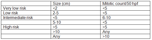
The behavior of these tumors is related to its site, with tumors in the small bowel having the worst behavior. Features that favor malignancy include the following: size larger than 5cm, necrosis, hemorrhage, hyper-cellularity, nuclear atypia, and mitotic activity (mitotic count greater than 5 per 50 HPF).
(Adapted from Gastrointestinal and liver pathology - Foundations in diagnostic pathology, 2005)
In very rare cases, multiple GISTs may be detected in one or more organs - not necessarily an indicator of greater aggressivity. However, the prognosis and treatment may differ from those of conventional GISTs. Multiple tumors may arise in three clinical contexts: sporadic tumor formation, familial GIST syndrome (autosomal dominant inheritance), or as an additional component of certain syndromes (Carney's triad, Carney-Stratakis syndrome and type I neurofibromatosis). The differential diagnosis of these syndromes is based mainly on clinical and genetic studies, rather than on morphological, immunohistochemical or molecular findings.
Immunohistochemical features include positivity for CD117(KIT) (95%), CD34 (60-70%), smooth muscle actin (30-40%), S-100 protein (5%), and more rarely desmin (1-2%). Two new tissue markers (DOG-1, PKC-theta) have been identified, providing specificity and sensitivity equal to or greater than those of CD117. Both proteins are selectively expressed on interstitial cells of Cajal (ICC), but their relationship with the KIT receptor is unknown. Molecular studies have revealed mutually exclusive mutations in KIT (60-80%) and PGFRA (5-10%) genes.
GISTs show universal expression of the receptor tyrosine kinase KIT. It is also established that mutations in KIT, or platelet-derived growth factor receptor-alpha (PDGFRA) lead to constitutive activation of their kinase activities, resulting in the development of GISTs. Imatinib (STI571) is a small molecule inhibitor of both kinases. This gene product-targeted therapy confers remarkable clinical benefit for patients with advanced disease.
Histology & Cytomorphology of GISTs
At histological examination, tumors tend to display spindle cell, epithelioid or mixed morphology.
The cytologic appearance depends on the cell type. The specimens are usually cellular, with isolated cells and loose and crowded fragments of spindle or epithelioid cells. The individual cells often lose their wispy cytoplasm and become stripped nuclei. Perinuclear or paranuclear vacuoles are present in some cells. Delicate cytoplasm and prominent nuclear palisading have also been noted.
Differential Diagnoses
The differential diagnosis depends on the primary cell types.
GISTs comprised of bland spindle cells should be differentiated from smooth muscle tumors (eg. leiomyoma), fibromatosis, solitary fibrous tumor, inflammatory myofibroblastic tumor and neural tumors (eg. Schwannoma), etc.
GISTs comprised of malignant spindle cells should be differentiated from leiomyosarcoma, malignant fibrous histiocytoma and dedifferentiated liposarcoma, etc..However, cytology in these tumors are much more pleomorphism than that in GISTs (even malignant lesions), and immunohistochemistry for CD117, CD34 and DOG1 are all negative.
GISTs comprised of epithelioid cells should be differentiated from poorly differentiated carcinoma, melanoma or clear cell sarcoma, glomus tumor, gangliocytic paraganglioma, endocrine carcinoma and benign epithelioid nerve sheath tumor, etc..
REFERENCES
![]() Contributed by Lin Liu, MD, PhD and Sara E Monaco, MD
Contributed by Lin Liu, MD, PhD and Sara E Monaco, MD