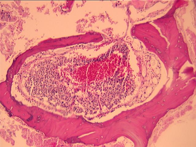
 H & E (1995)
MICROSCOPIC DESCRIPTION:
H & E (1995)
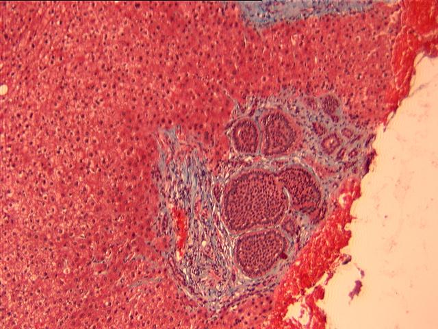
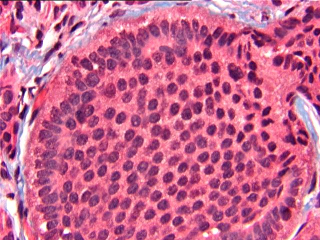 Trichrome (1989)
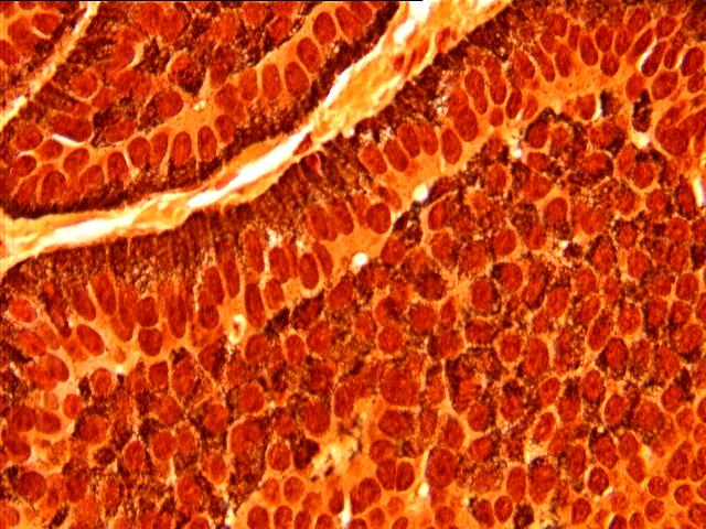 Grimelius (1989)
Sections show multiple fragments of fibrous tissue and bone with focal cautery and crush artifact. In part 1 there are foci of crushed hyperchromatic clusters of small, blue cells, probably representing the metastatic carcinoid. Parts 2, 3, and 4 show more extensive areas of well preserved metastatic carcinoid tumor, focally filling the bony trabeculae.
Grimelius stain from metastatic carcinoid in 1989 reveals neurosecretory granules.