)
)
)
)
)
)
)
IMMEDIATE IMPRESSION
MASS, COCCYGEAL, FINE NEEDLE ASPIRATION BIOPSY, PERFORMED BY PATHOLOGIST -
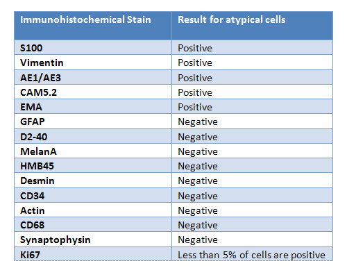
The aspirates reveal fragments of metachromatic matrix and mildly atypical epithelioid cells. These cells are predominantly arranged in nests and cords, though occasional isolated cells are within the background of the matrix material are noted. Of note, the cells have abundant amounts of vacuolated cytoplasm (see image 3).
Given that mucinous, chondroid and myxoid lesions may display similar cytomorphologic features, a combination of clinical, radiologic, morphologic and immunohistochemical clues are needed to arrive at a final diagnosis.
The next day, the Papanicolaou-stained slides and cell block are ready (images 5 and 6). Immunohistochemical stains are ordered, and the results are also shown below (images 7, 8, 9 and 10).