

MICROSCOPIC DESCRIPTION:
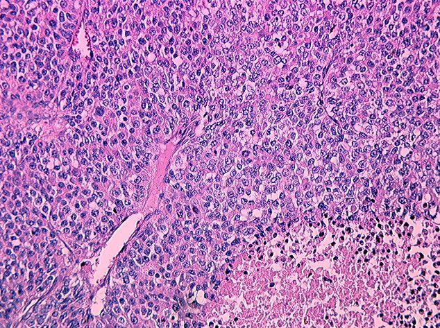
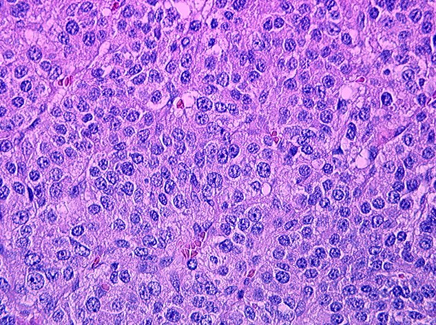
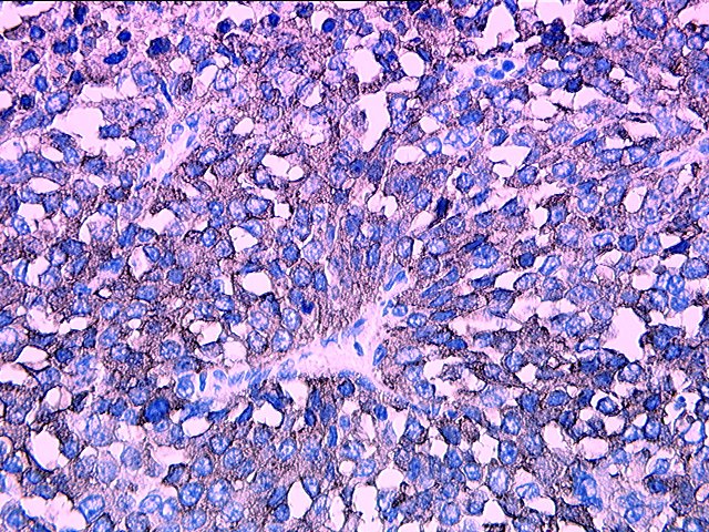
The resected neoplasm was confined to the adrenal gland. At the edges, infiltration through the capsule of the adrenal gland was noted. The neoplasm consisted of sheets of polygonal cells with a rich vascular stroma and areas of geographic necrosis. Cytologically, the vast majority of the cells were polygonal with eosinophilic cytoplasm, a moderate degree of nuclear pleomorphism, prominent nucleoli, and vesicular chromatin pattern. In the most active areas, 35 mitoses were counted in 10 high powered fields. Invasion into the adrenal vein seen grossly was confirmed histologically. In an attempt to confirm the adrenal cortical origin of this neoplasm, a battery of immunohistochemical stains were performed. Intense staining for Synaptophysin was appreciated. Vimentin stain was also positive. Immunohistochemical stains for AE1/AE3 (cytokeratin), CAM 5.2, Epithelial Membrane Antigen, and Chromogranin were negative.