


MICROSCOPIC DESCRIPTION:
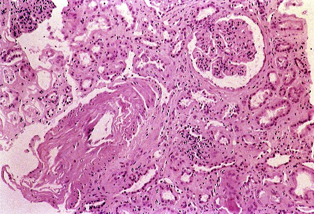
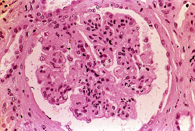
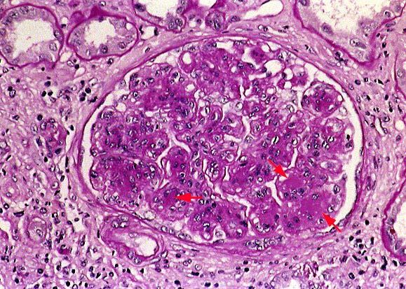
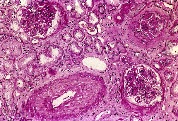
Tissue examined for light microscopy consists predominantly of renal cortex including renal capsule and a small amount of medulla. The profiles of up to 46 glomeruli are identified in the paraffin, frozen, and plastic embedded tissue, of which 12 (26%) are globally sclerotic. Most of the sclerotic glomeruli are concentrated in one core in a subcapsular location. The non-sclerotic glomeruli show a diffuse type I membranoproliferative pattern of injury with prominent intracapillary neutrophils and basement membrane reduplication (tram tracking, red arrow) noted on the silver stain. In addition, PAS positive hyalinized foci (red arrows) are present in several mesangial regions. No necrotizing lesions or crescents are seen.
The tubules show significant patchy but widespread atrophy and focal acute tubular injury. Some of the tubules contain oxalate crystals, rare RBC casts, and occasional pigmented (? hemoglobin) granular casts. The interstitium is expanded by moderate fibrosis corresponding to the areas of tubular atrophy.
There is patchy, chronic and focally acute interstitial inflammation. Scattered neutrophils are concentrated focally in the distribution of peritubular capillaries suggesting the possibility of an acute peritubular capillaritis. The larger intrarenal arteries show prominent fibroelastic intimal hyperplasia (example 1 and example 2) with marked luminal narrowing. The arterioles show myointimal thickening. No arteritis/arteriolitis nor acute sclerodermal vascular changes / onion skinning is seen. The juxtaglomerular apparati are not prominent.
IMMUNOFLUORESCENCE:
RESULTS:
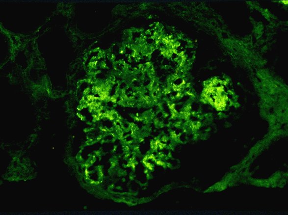
The tissue examined for immunofluorescence contains eight lobular, non-sclerotic glomeruli, two arteries and five arterioles. Direct immunofluorescence is performed using a panel of ten antisera.
| . | IgG | IgM | IgA | Clq | C3 | C4 | Fib | Alb | KAP | LAM |
|---|---|---|---|---|---|---|---|---|---|---|
| Glomeruli: Linear diffuse |
- | - | - | - | - | - | - | 2+ | - | - | Granular (GBM) | 3+ | 2+ | - | - | 3+ | - | - | - | - | 2-3+ (focal) |
Granular (MES) | 3+ | - | - | 1+ | 3+ | - | - | - | TR | 3+ (focal) |
Focal | - | - | - | - | - | 1-2+ | **3+ (Urinary Space) | - | - | - | Other | - | - | 2+* | - | - | 2+ (Urinary Space) | - | 2+ (Bowman's Capsule) | - | - | Tubules: Basement Membrane |
- | - | - | - | - | - | - | 2-3+ | - | - | Casts | - | + | - | - | - | - | - | - | - | - | Interstitium: Connective Tissue |
2+ | - | - | - | - | - | - | - | 2+ | 1+ | Vessels: Capillary |
- | - | - | - | - | - | 2+ | - | - | - |
*Rare visceral epithelial staining; **Irregular GBM and
Abbreviations: Fib - Fibrinogen; Alb - Albumin; KAP - Kappa; LAM - Lambda; GBM - Glomerular basement membrane; MES - Mesangium; M - Mural; I - Intimal
INTERPRETATION:
IMMUNE COMPLEX GLOMERULONEPHRITIS, MEMBRANOPROLIFERATIVE PATTERN, WITH IgG, IgM, C1q, C3 AND LAMBDA LIGHT CHAIN-RESTRICTED STAINING.
ELECTRON MICROSCOPY:
RESULTS:
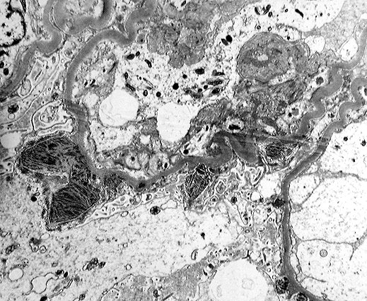
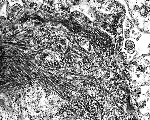
A single glomerulus showing a membranoproliferative pattern of injury is examined ultrastructurally. The podocytes are swollen, with rare foot process fusion and slight microvillous transformation noted. In many areas of the glomerulus, there is prominent mesangialization of the capillary loops characterized by mesangial interposition and reduplication of the subendothelial basement membrane, with electron dense fibrillary material present predominantly in mesangial areas and the mesangialized subendothelial space. Some fibrillary material irregularly distributed as large hump-like deposits is present in the subepithelial space extending into the urinary space. The fibrils in some locations are oriented in organized parallel configurations and in other areas are more haphazardly arranged. The fibrils range in diameter from approximately 15 to 50 nm and have a vaguely microtubular appearance. At higher magnification, each of the fibrils has a very regular crystalline substructure, morphologically consistent with immunotactoid. Between the fibrils, there is amorphous electron dense material. One capillary loop contains an intraluminal neutrophil. Many of the endothelial cells are swollen. No tubuloreticular inclusions or basement membrane breaks are identified. Several tubules examined show degenerative epithelial and basement membrane changes. No immunotactoid is present outside of the glomerulus.
INTERPRETATION:
TYPE I MEMBRANOPROLIFERATIVE GLOMERULAR PATTERN OF INJURY DUE TO IMMUNOTACTOID DEPOSITION.