![]() Contributed by by Shveta Hooda, MD, Ken Ho, MD and William Pasculle, ScD
Contributed by by Shveta Hooda, MD, Ken Ho, MD and William Pasculle, ScD
PATIENT HISTORY:
A fifty-four year old female with past medical history of pulmonary hypertension, fibromyalgia, chronic obstructive pulmonary disease, coronary artery disease and hypertension presented to the emergency department with complaints of increased dyspnea, lower extremity edema, weight gain, orthopnea, fevers, chills, night sweats over the past few months. She denied any cough or sputum production. The patient was on trepostinil (remodulin) infusion since 3 years. She had a history of line infection two years ago which was successfully treated with antibiotics. Patient had a right subclavian port placed about ten months ago. Her other medications included estrogen, escitalopram, warfarin and gabapentin. The patient did have a remote history of smoking although she had quit smoking eight years prior. She denied any drug allergies, alcohol or intravenous drug abuse.
On presentation her vitals were found to be a blood pressure of 160/90, heart rate 88, temperature 36.9, and oxygen saturation 93% on 2 liters by nasal canula. The review of systems was essentially negative except for the history of present illness. Her examination was positive for tachypnea, a 3/6 systolic murmur, 2+ bilateral lower extremity pedal edema. Rest of the examination was normal.
Blood cultures were drawn in the emergency department. Initial lab evaluation showed WBC count 8700 / mm3, neutrophils 83%, lymphocytes 13% and hemoglobin 13.3 g/dl. Electrolytes, BUN and creatinine were normal.
)
)
)
)
)
)
Patient underwent transthoracic echocardiogram which showed a small mobile mass (vegetation) on the aortic side of the aortic valve (Figure 1) and another round mass in the right ventricle attached to chordae or the moderator band measuring 1.2 cm in diameter.
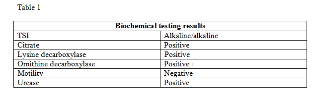
The blood cultures grew plump gram negative coccoid rods (Figure 2) on day 3. The blood culture isolate grew pale-pink shiny raised and mucoid colonies on 5% sheep blood agar plates (Figures 3, 4 and 5). The biochemical results (Figure: 6) were as follows (Table 1):
The Microscan identified the bacteria as Oligella Urealytica (67.2%). APINFT was done which again identified the bacteria as Oligella Urealytica (89%).
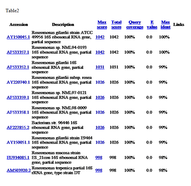
As both of the above tests did not give good identification of the bacteria 16S DNA sequencing (Table 2) was done to identify it.