MICROSCOPIC DESCRIPTION:
Thyroid
Lymph Node

Immunoperoxidase --
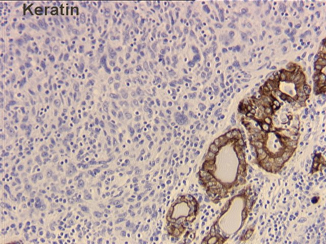
Keratin,
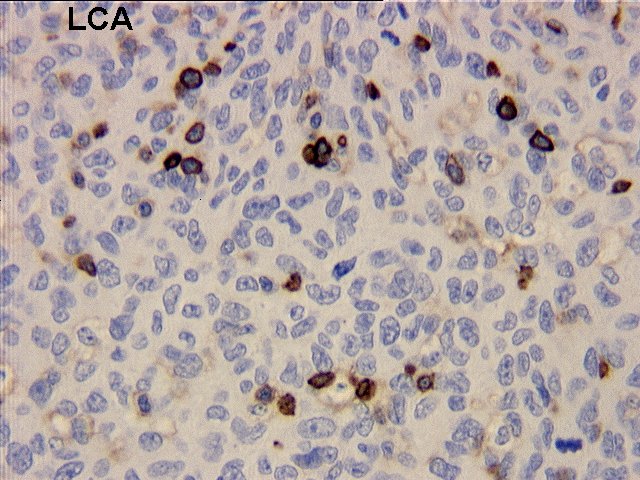
LCA,
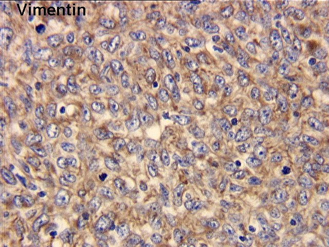
Vimentin,
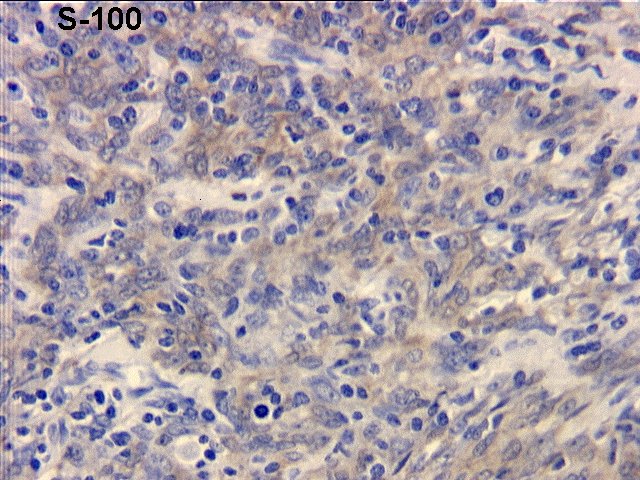
S100,
CD21,
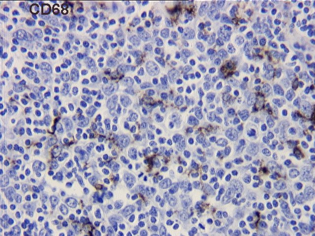
CD68
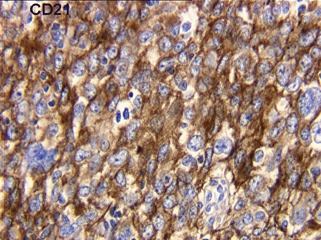
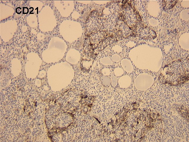
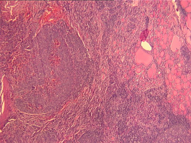
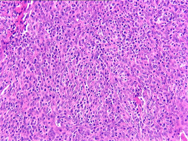
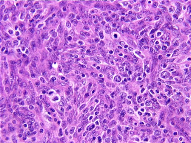
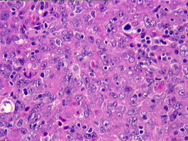
Microscopic examination reveals a spindle cell tumor with interspersed lymphocytes growing in a nodular pattern and totally replacing the normal architecture of one of the lymph nodes from the right neck. Additionally, the same proliferation of spindle cells, with large nuclei, prominent nucleoli and a moderate amount of cytoplasm, is present as a large mass within the thyroid parenchyma, as well as in the lymphoid tissue of the thyroid gland. The tumor appears to be growing in nodules and sheets, with occasional whirling and storiform patterns. Mitotic figures are readily identifiable.
| STAIN | RESULT |
|---|---|
| Keratin | Negative |
| LCA | Negative |
| Vimentin | Positive |
| S-100 | Weak Positive |
| CD21 | Positive |
| CD68 | Focal Positive |