
MICROSCOPIC DESCRIPTION:
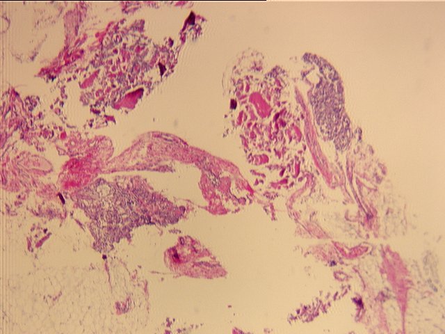
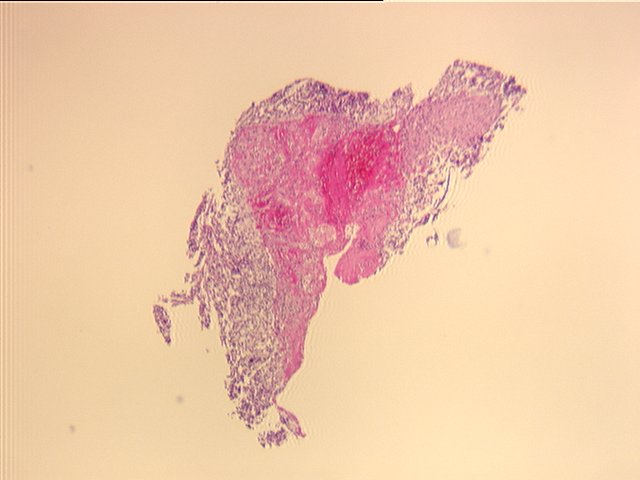
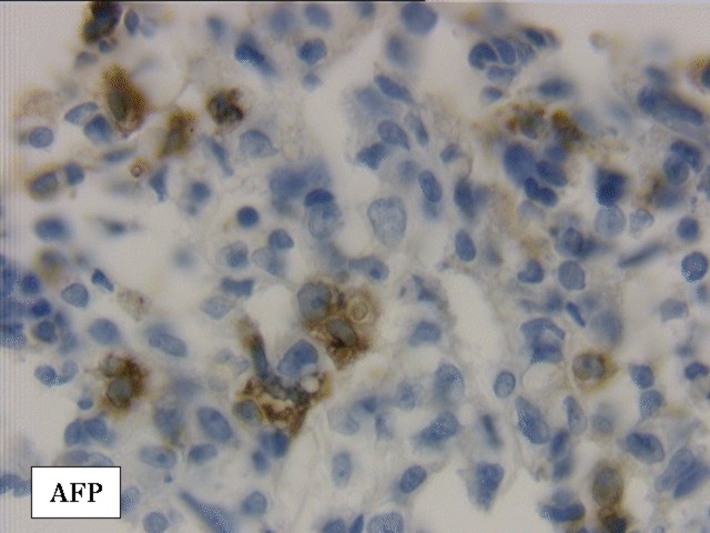
Sections show pieces of skeletal muscle, bone and adipose tissue with small fragments of tumor. The tumor is composed of numerous cells with round-oval hyperchromatic nuclei many with prominent nucleoli and varying amounts of eosinophilic cytoplasm. Some cells have one or more round hyaline droplets in their cytoplasm. These hyaline droplets are PAS-positive and resistant to diastase digestion. Immunohistochemical stains for PGP are negative with high background. Desmin is negative and scattered cells are strongly positive for alpha-fetoprotein (AFP).