
GROSS DESCRIPTION:
The specimen is received in five parts.
Part 1:
Parts 1 through 4 consist of regional lymph nodes ranging from 0.3 to 2.0 cm. They are entirely submitted.
Part 5:
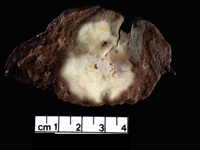
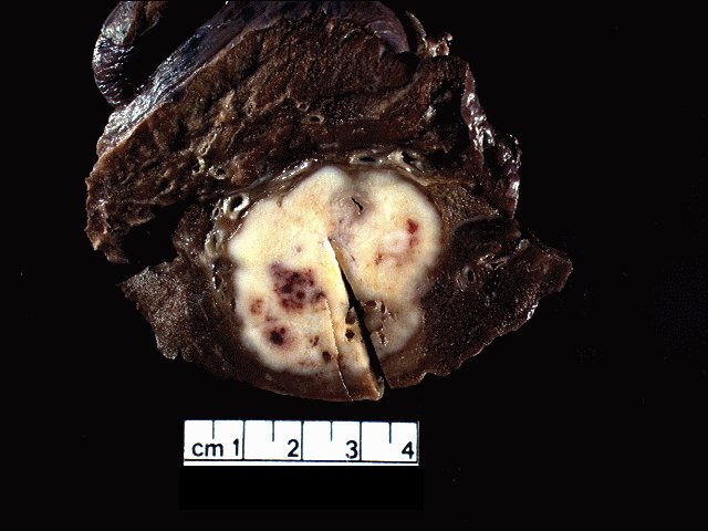
Part 5 is received unfixed labeled "right upper lobe mass", and consists of a 9.5 x 9.0 x 6.5 cm., 150 gram (fixed weight), resection of lung. The visceral pleural surface is smooth, tan- purple and focally anthracotic with stapled resection margins. There is a focal, 2.5 x 0.7 cm. area of visceral pleural puckering. The bronchial and vascular resection margins have been previously removed for frozen section evaluation. Following frozen section evaluation, the bronchial and vascular resection margins are entirely submitted in cassette 5A. Multiple anthracotic peribronchiolar lymph nodes are dissected, the largest of which is 1.0 cm., and submitted in cassette 5B. The stapled resection margins are removed and the previous site of the staples are inked. Upon sectioning, a 4.8 x 4.0 x 4.0 cm firm, tan, focally hemorrhagic and necrotic mass is seen underlying the area of pleural puckering. The mass comes to within 0.5 cm. of the closest stapled resection margin. The surrounding normal lung parenchyma is tan-red and spongy without evidence of other lesions. Tissue is taken and frozen in OCT and bulk, and for DNA for tissue bank. Representative sections are taken and submitted as follows: 5C - mass and closest stapled resection margin 5D - mass adjacent to puckered pleural surface 5E - tumor and normal stent 5F - normal lung.
INTRAOPERATIVE CONSULTATION: 5A: LUNG, RIGHT UPPER LOBE, BRONCHIAL AND VASCULAR RESECTION MARGINS, RESECTION - BRONCHIAL AND VASCULAR RESECTION MARGINS ARE FREE OF TUMOR.