
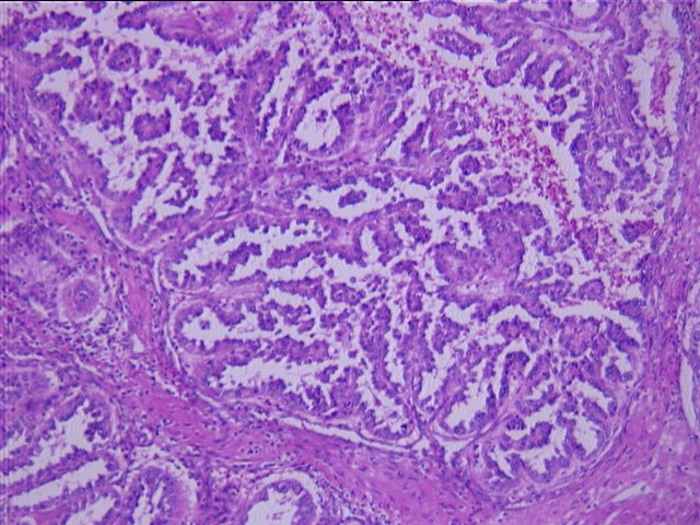
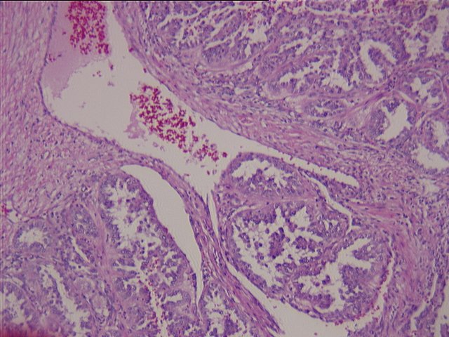

MICROSCOPIC DESCRIPTION:
The lobular neoplastic growth pattern is well circumscribed by a desmoplastic response. Large areas of tumor necrosis are present which in some areas results in cystic change. Within the lobules of the tumor are well formed papillae with fibrovascular stalks supporting the malignant cells. Foamy macrophages infiltrate the fibrovascular cores focally, however cystic spaces show areas of aggregated phagocytes in response to epithelial sloughing. Focally, small vessels within the tumor exhibit invasion by malignant cells. The malignant cells are characterized by polygonal vesicular nuclei allowing visualization of prominent nucleoli.