GROSS DESCRIPTION:
The specimen is received in four parts.
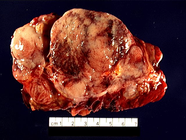
Part 1:
Part 1 is received as part of an intraoperative consultation
labeled "mediastinal mass". It consists of a 16.0 x 10.0 x 5.0
cm portion of soft tissue with an attached 2.0 x 1.0 x 1.0 cm
portion of lung closed by staples. There is also an attached 7.5 x 5.0 x
0.2 cm portion of tan-purple, smooth and glistening tissue, consistent with
pericardium. The surface of the specimen contains a moderate
amount of yellow, lobulated unremarkable fat with strands of red-
brown unremarkable muscle. Upon sectioning, there is an 11.0 x
7.2 x 3.0 cm well-circumscribed, yellow-tan, mass with an apparent
pseudocapsule. This mass is actually separated into three
discrete tan, firm, focally hemmorrhagic and calcified lobules, up to 8.5 cm in greatest dimension, separated by connective tissue and a small
amount of fat. There are also occasional cystic spaces up to 0.2 cm containing serous fluid.
Part 2:
Part 2 is labeled "superior left thymic extension". It consists
of a two tan, firm, round nodules, 2.5 and 2.0 cm respectively, with attached fat. The surfaces of the nodules are tan, smooth and glistening. Upon sectioning, both appear to have a
pseudocapsule, are tan, soft and homogeneous.
Part 3:
Part 3 is received in normal saline labeled "normal thymus". It
consists of 11.8 grams, 5.2 x 4.0 x 1.2 cm of tan, red-brown,
soft tissue, consisting of fat, muscle, small lymph nodes and a
0.5 cm tan, homogeneous well circumscribed nodule.
Part 4:
Part 4 is received as part of an intraoperative consultation labeled
"parathyroid". It consists of a 0.097 gram, 0.8 x 0.7 x 0.6 cm portion of parathyroid gland. The portion received in pathology is only one-third of a larger ectopic parathyroid gland found in the left thoracic outlet during surgery.