Electron Microscopy -- Left Inguinal Lymphadenopathy
ELECTRON MICROSCOPY:
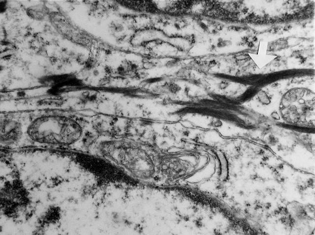
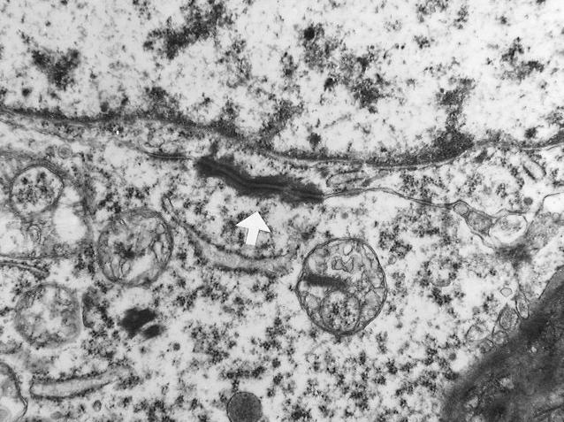
Electron microscopy reveals numerous desmosomes and abundant tonofilaments. No evidence of melanocytic differentiation, adenocarcinoma differentiation nor sarcomatoid differentiation is seen.