


MICROSCOPIC DESCRIPTION:
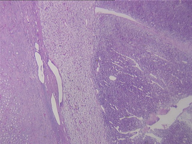
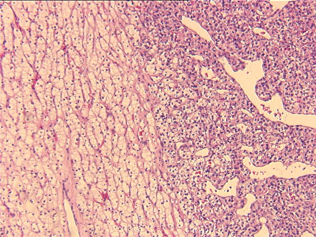
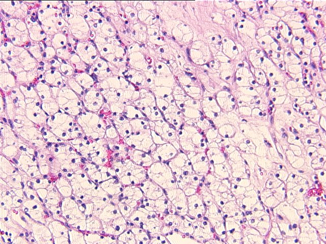
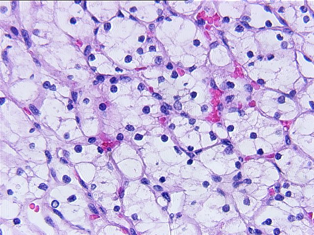
Slide 1 and slide 2 show a renal mass which is composed of two distinct components surrounded by a mature collagenous capsule and a rim of compressed renal tissue. The border between each cell type is distinct. Slide 3 and slide 4 show the peripheral zone of the mass which consists of large, polygonal cells with clear cytoplasm arranged in an alveolar growth pattern with uniform, round nuclei and inconspicuous nucleoli. Slide 5 and slide 6 show the central zone of the mass which consists of fairly uniform epithelioid cells arranged in a trabecular growth pattern and surrounded by a rich vascular network. At low magnification, these elongated, branching blood vessels confer a hemangiopericytoma-like growth pattern upon the tumor. At higher magnification, slightly pleomorphic nuclei are seen which are surrounded by a finely granular cytoplasm. No mitoses are observed. Slide 7 shows diffuse hyperplasia of the zona glomerulosa of the adrenal gland.
ELECTRON MICROSCOPIC DESCRIPTION:
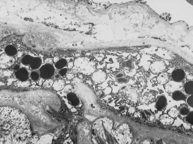
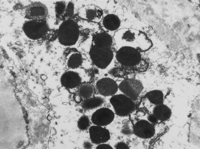
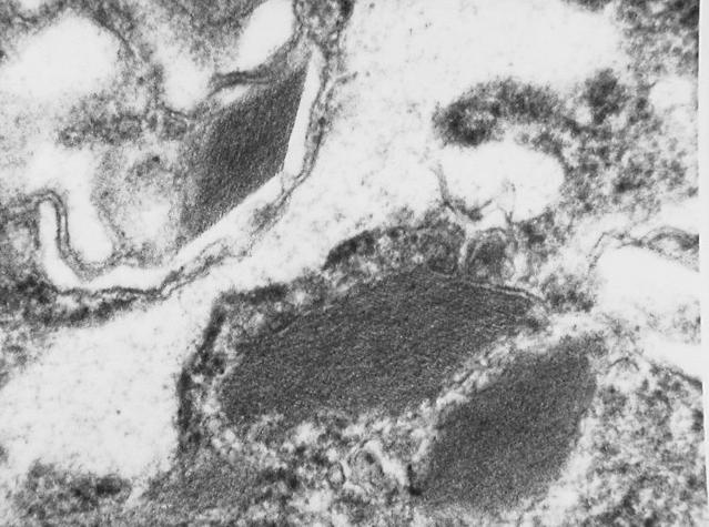
Slide 8, slide 9, and slide 10 are electron micrographs taken from the central zone of the mass showing the characteristic ultrastructural intracytoplasmic granules of a juxtaglomerular tumor. Two types of granules are seen. The more frequent granules are round and electron dense and represent mature renin. The less frequent granules are sharply angulated with a periodic crystalline substructure and represent the immature protogranules of renin.