

MICROSCOPIC DESCRIPTION:
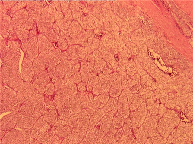
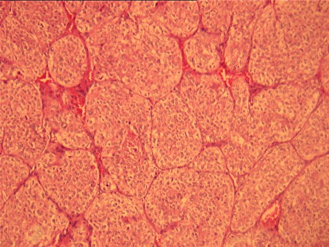
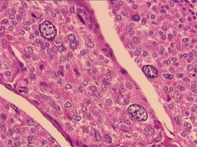
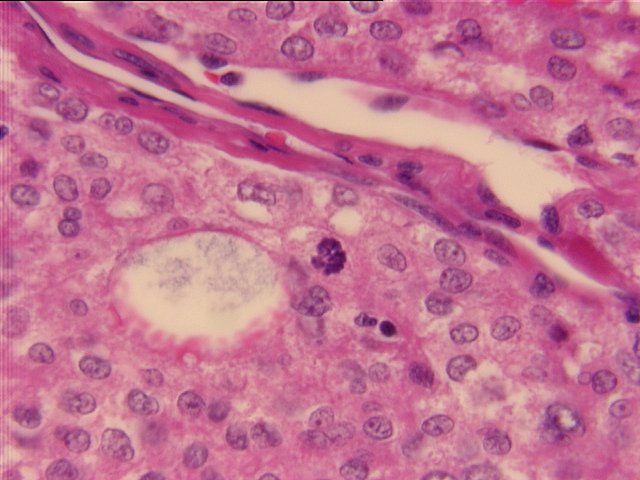
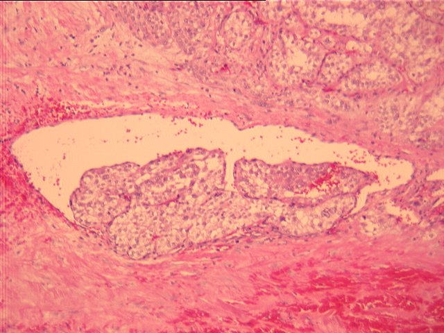
Microscopic examination reveals an encapsulated mass comprised of nests of cells separated by a fine fibrovascular septa. The cells are relatively monomorphic, with abundant pink granular or fibrillary cytoplasm, indistinct cytoplasmic borders, and relatively stippled occasionally vesicular nuclei. Infrequent pleomorphic cells are present. Nucleoli and mitotic figures are not prominent. Vascular invasion is noted. Extension of the tumor past the capsule is seen.
A congo Red stain for amyloid deposition was negative. A mucicarmine stain was positive. Immunoperoxidase stain results were negative for thyroglobulin, positive for prekeratin, negative for chromogranin, negative for calcitonin, and negative for the cytokeratins AE1 and AE3.