
 PAS/D
PAS/D
MICROSCOPIC DESCRIPTION:
PAS/D
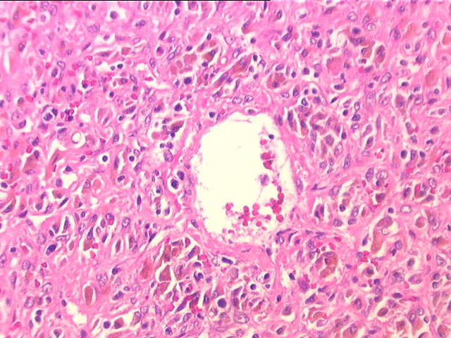
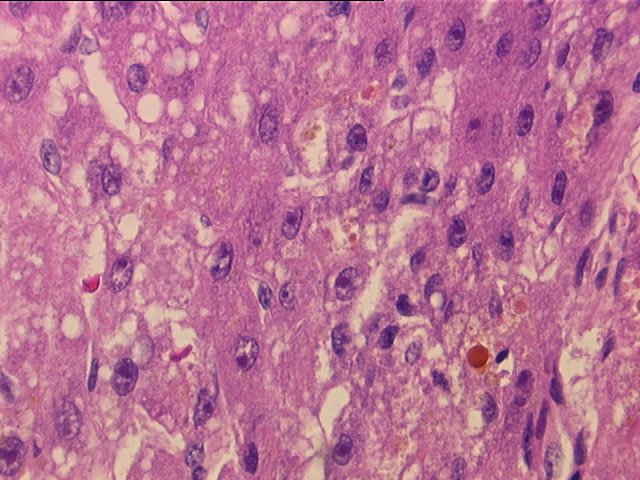
Sections through the liver show distorted hepatic architecture, due to inflammatory bridging (central-to-central)necrosis. The viable hepatocytes are located in the periportal areas. Mild cholangiolar proliferation is seen. The portal tracts contain a mild lymphoplasmacytic infiltrate enriched in areas with eosinophils. The most striking histopathologic changes are in the centrilobular areas. There is hepatocellular dropout and hemorrhage with hemosiderin latent macrophages and mild lymphocytic infiltrate with focal central vein endothelialitis. A few central veins show pericentral fibrosis. The viable hepatocytes demonstrate regenerative changes, mild hepatocellular swelling, cholestasis, and moderate micro- and macrovesicular steatosis. No viral inclusions are appreciated. The PAS-D stain is negative for alpha-1 antitrypsin globules. In section 1D, from the superficial hilum, the hepatic artery demonstrates mild fibro- intimal hyperplasia. The common hepatic duct shows mild chronic mural inflammation and the periductal glands demonstrate mild reactive changes. The gallbladder shows mucosal autolysis and mild mural thickening. The hepatic vein resection margins are unremarkable. The grossly described regenerative nodules (sections 1H and 1I), show more prominent cholestasis and macrovesicular steatosis. Overall, the changes are suggestive of a drug induced injury, rather than a viral infection and they are consistent with the patient's clinical history of tylenol overdose.