
GROSS DESCRIPTION:
The specimen is received in three parts:
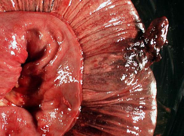
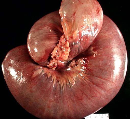
PART 1 consisted of a knotted mass of small bowel having a grossly dark red necrotic appearance. Two stapled surgical margins were visible on the specimen. The first margin consisted of a 4.5 cm length of small bowel which was light tan and viable in appearance. The opposite margin consisted of a 27 cm length of dusky red necrotic-appearing bowel.
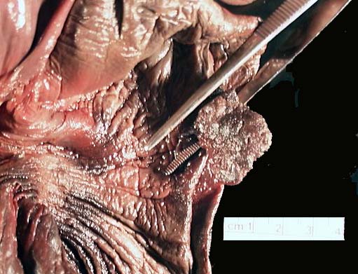
The two stapled ends were opened, revealing grossly viable mucosa at the first margin and dusky red, grossly necrotic mucosa at the second margin. The specimen was unknotted/reduced revealing the central portion of the specimen to consist of 93 cm of intussuscepted bowel having a dusky red, necrotic appearance with no apparent perforations. The specimen was opened, revealing two mucosal polyps.
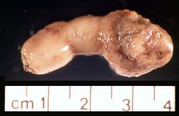
The first polyp measures 5.0 x 4.5 x 2 cm with stalk measuring 2 cm in length and 1 cm in diameter; it is located at the edge of the intussuscepted region, 6 cm from margin 1. It had a red, beefy, shaggy verrucous appearance reminiscent of a adenomatous polyp; it had no apparent internal structure. The second polyp measures, 4 x 2.5 x 1 cm and was found on the mucosa 34 cm from margin 2 (within the intussuscepted region). It had a firm, slightly lobulated, solid consistency similar to that of a blood clot.