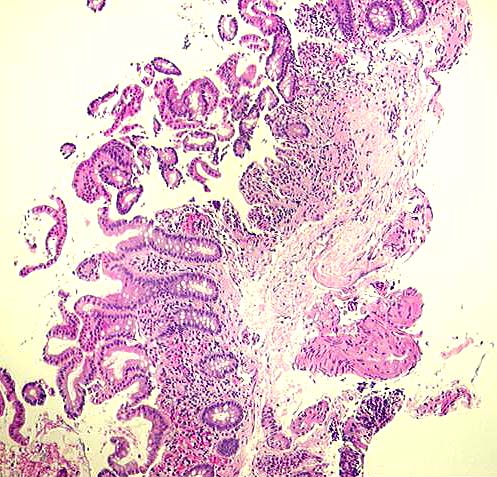

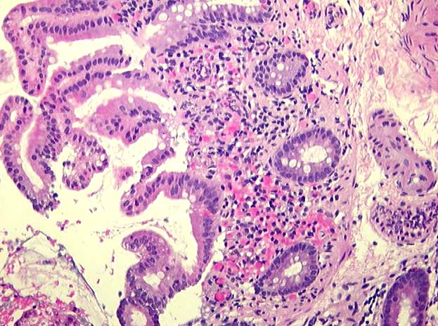
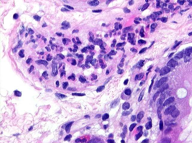
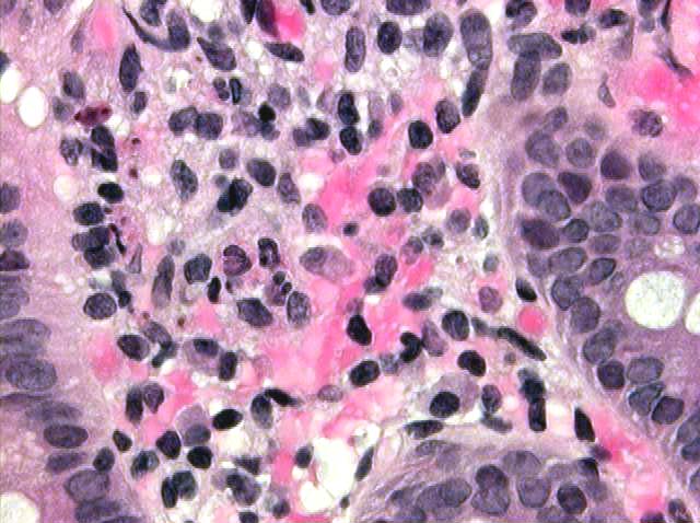
MICROSCOPIC DESCRIPTION:
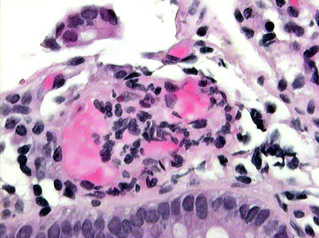
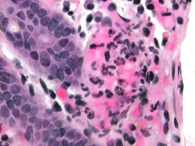
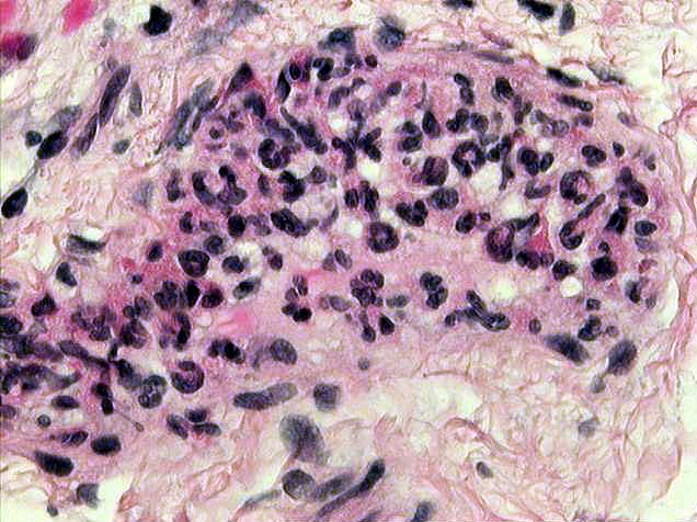
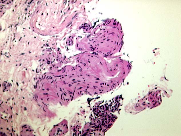
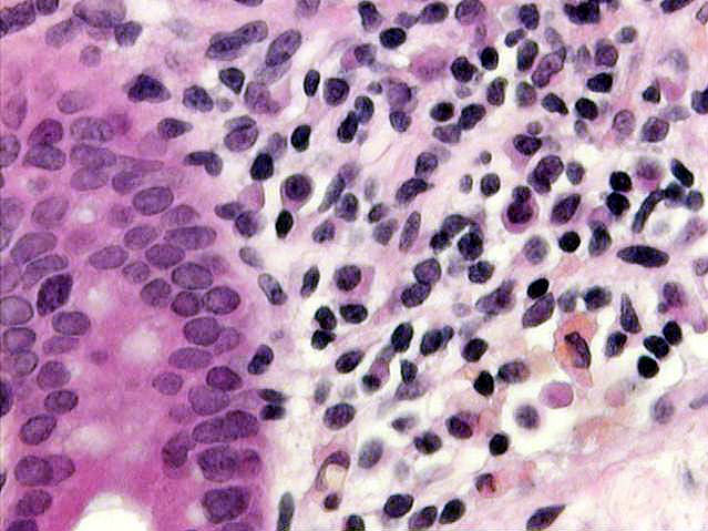
The biopsy specimen consists of a portion of cecal mucosa with a portion of underlying submucosa with several larger submucosal vessels. Examination of the surface of the biopsy demonstrates colonic epithelium with goblet cells and moderately distorted crypts. Some areas of the lamina propria are expanded by a moderate number of lymphoid cells with admixed plasma cells. Other areas of the lamina propria demonstrate focal hemorrhage, and collections of neutrophils and probable fragments of nuclear debris. Examination of the submucosa reveals edema and numerous small vessels which display varying amounts of neutrophilic infiltration and destruction of the vascular wall. Some of the vessels demonstrate tortuosity and occlusion by a combination of neutrophils, thrombosis, nuclear debris, and eosinophilic necrosis of the vessel wall. In the deepest portion of the biopsy, a thickened, tortuous, moderately fibrotic vessel is noted, suggesting a chronic vasculitic/inflammatory process.