

MICROSCOPIC DESCRIPTION:
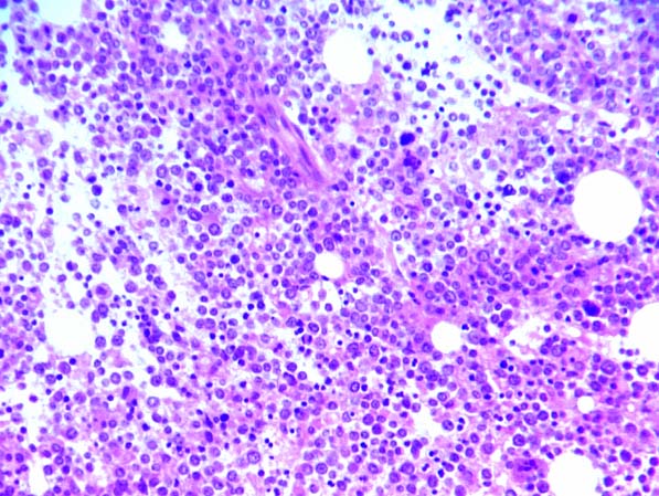
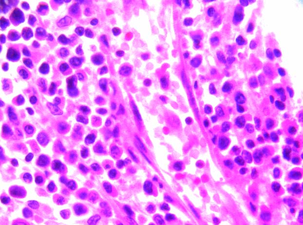
The bone marrow demonstrated a homogeneous infiltrate composed of intermediate size mononuclear cells with prominent nucleoli and plasmacytic differentiation admixed with small lymphocytes. The remainder of the tissue was normocellular with some areas of trilineage hematopoiesis, although the erythroids appeared slightly decreased.
Immunohistochemistry on paraffin identified a monoclonal population of B-cells with CD20/L26 positive, anti-lambda positive, CD3 negative and CD5 negative phenotype.
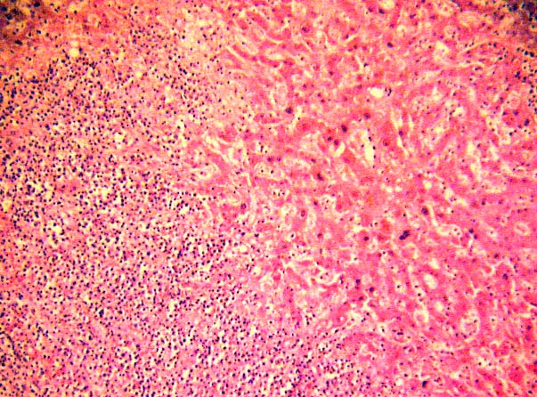
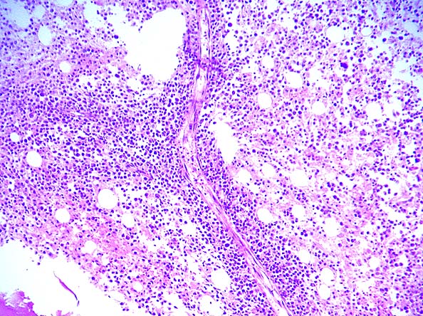
Antemortem cytometric immunophenotypic studies performed on the bone marrow supported the findings of a malignant B-cell neoplasm. In addition, there was a relative increased number of CD3 negative NK cells which were CD16, CD56 and CD7 positive. Some of the cells were CD57 positive as well. These cells did not express CD2. The significance of the smaller population of NK-type cells is unclear. Postmortem immunohistochemical stains preformed on the bone marrow and liver and supported the antemortem diagnosis of a malignant B-cell neoplasm as well as an accompanying lymphocytic population with CD2 positive, CD3 negative CD79a positive phenotype. The normal architecture of the liver was replaced by large geographic areas of bridging necrosis. There was a neoplastic lymphoid infiltrate involving the periportal, central lobular and sinusoidal areas. There was no distinct pattern of infiltration. The sinusoids were expanded with hemorrhage, edema and neoplastic cells. Viable hepatocytes showed regenerative changes. There was no fibrosis, viral inclusions and no evidence of primary biliary tract pathology.
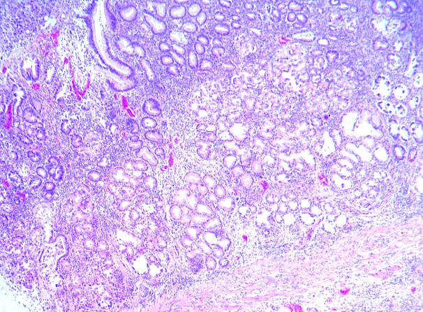
The normal architecture of the spleen was effaced with multiple infarcts and a diffuse lymphoid infiltrate, involving the white and red pulps as well as diffuse infiltration of the sinusoids. There was focal extramedullary hematopoiesis.
Microscopic examination of the lymph nodes revealed rare atypical lymphoid cells in predominantly necrotic tissue.