


MICROSCOPIC DESCRIPTION:
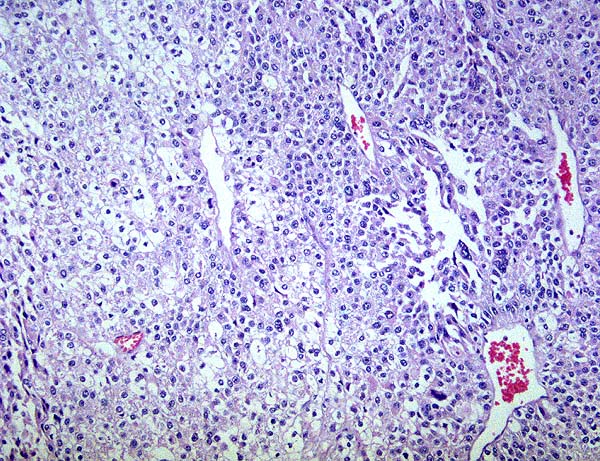
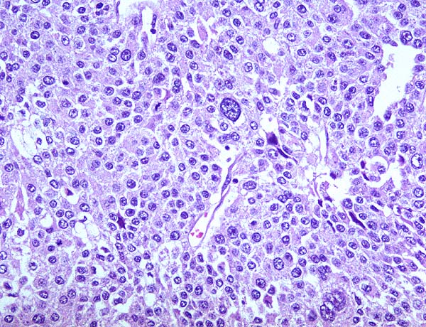
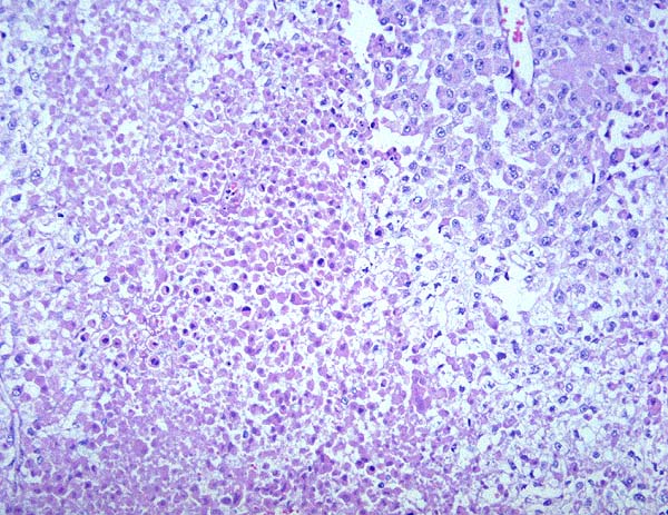
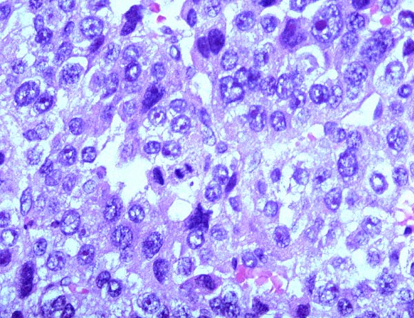
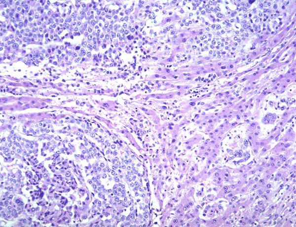
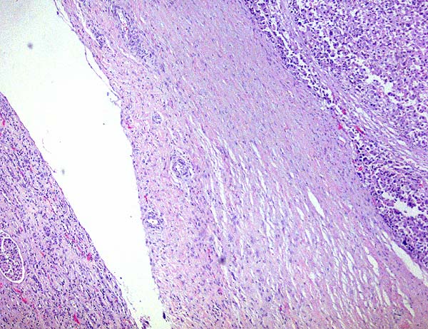
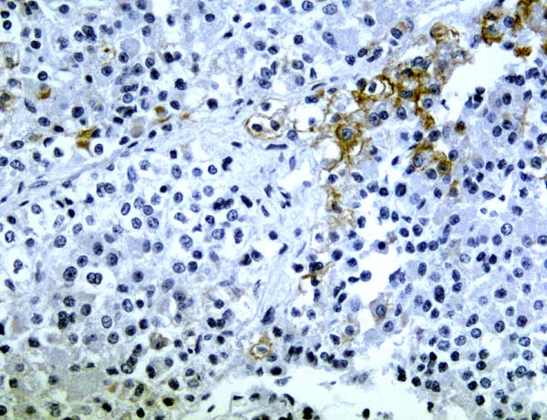
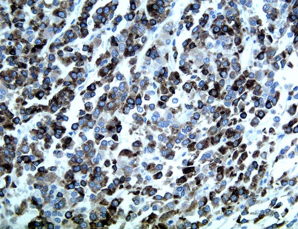
The tumor is composed of vague nests of cells separated by a fine vascular network. The cells are somewhat polygonal but in areas have indistinct borders with clear to partially eosinophilic cytoplasm. The nuclei are occasionally large and irregular with an open chromatin pattern and occasional small nucleoli. Mitotic figures are easily identified. Areas of necrosis are admixed with viable tumor. Nests of tumor cells are identified within the hepatic parenchyma. A band of fibrous tissue is identified between the tumor and kidney with no microscopic invasion seen. PAS stain is focally positive but is sensitive to diastase. Immunohistochemical stains for low molecular weight cytokeratin (CAM 5.2) and wide spectrum cytokeratin are focally positive. Vimentin is strongly positive. Epithelial membrane antigen is negative. There is strong immunoreactivity for inhibin A.