Microscopic Description -- Dilated Cardiomyopathy
MICROSCOPIC DESCRIPTION:
HEART:
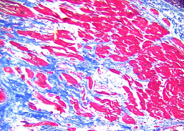
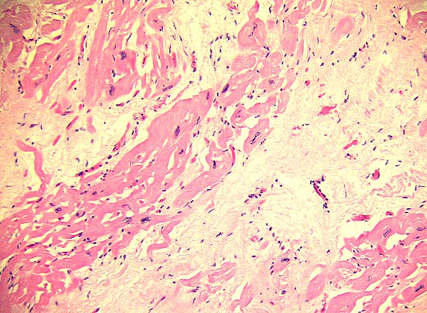
 The right and left atria showed moderate nuclear pleomorphism and marked endomysial fibrosis. The left and right ventricles and the interventricular septum showed severe nuclear pleomoiphism, marked endomysial fibrosis and marked cellular atrophy (Figures 3A and 3B). The right ventricle and the interventricular septum showed mild fatty infiltrate of the muscle wall.
LUNGS:
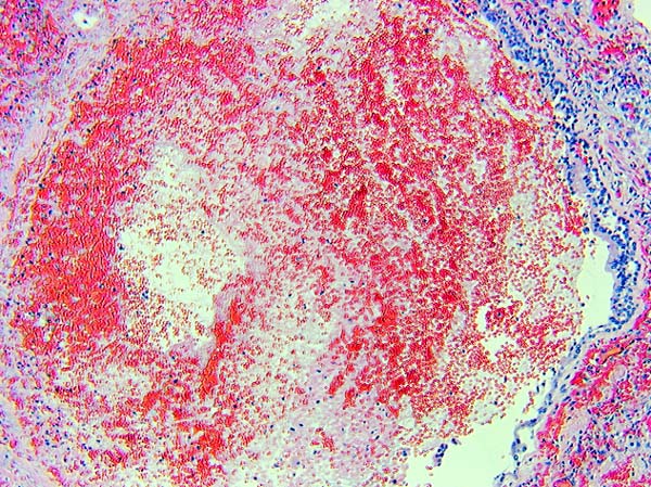
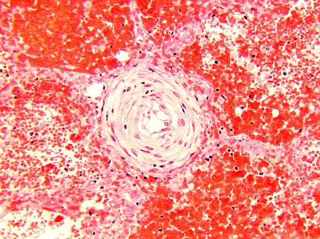
 The right lower lobe of lung showed severe alveolar hemorrhage with abundant hemosiderin laden macrophages, as well as intraalveolar capillary congestion and dilatation with focal septal fibrosis and necrosis (Figure 4). The smaller pulmonary arteries were thickened secondary to initial hyperplasia with myxoid change (Figure 5). Peribronchial vessels were dilated and congested. Numerous bronchioles contained fibrin thrombi and submucosal bronchiolar granulation tissue (Figure 6). The lung parenchyma showed focal atelectasis. Grocott and Gram stains were negative for organisms. The upper lobe of lung showed moderate atelectasis with numerous hemosiderin laden macrophages, mild alveolar hemorrhage and thickened pulmonary vessels, some of which contained fibrin thrombi. The left lung showed moderate atelectasis with numerous hemosiderin laden macrophages, mild alveolar hemorrhage, mildly thickened pulmonary vessels, marked subpleural vascular congestion and focal moderate alveolar capillary congestion and dilation.
SKELETAL MUSCLE:

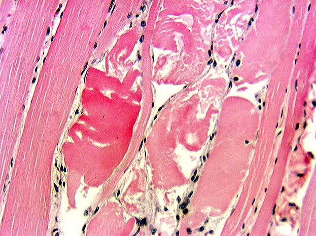
 Skeletal muscle from the right and left pectoralis muscles, intercostal muscle, right and left diaphragm and esophagus showed marked endomysial fibrosis with occasional fiber splitting, marked centralization of sarcolemmal fiber nuclei, fiber attenuation, varied fiber size, hypercontracted fibers, fiber regeneration and focal fiber necrosis (Figures 7A and 7B).
FINAL DIAGNOSIS


 The right and left atria showed moderate nuclear pleomorphism and marked endomysial fibrosis. The left and right ventricles and the interventricular septum showed severe nuclear pleomoiphism, marked endomysial fibrosis and marked cellular atrophy (Figures 3A and 3B). The right ventricle and the interventricular septum showed mild fatty infiltrate of the muscle wall.