IMAGING AND OPERATIVE DESCRIPTION:
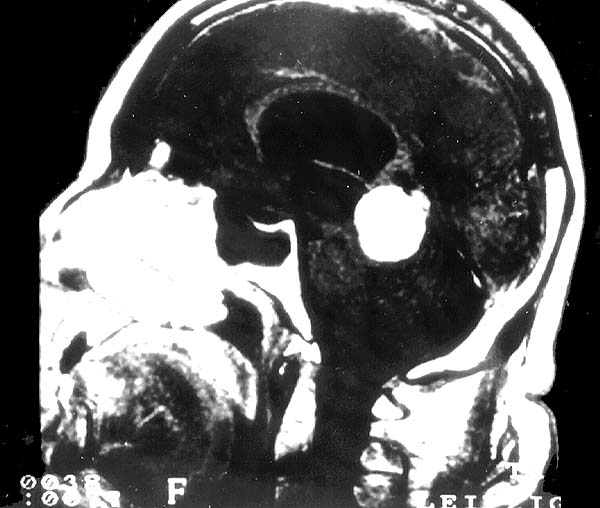
MRI studies revealed a clearly delimited tumor of the pineal region measuring 3.0 cm in diameter. The tumor was hyperintense by T2-weighting and enhanced with contrast medium. There was compression of the quadrigeminal plate and cerebellar vermis with stenosis of the aquaeduct and occlusive hydrocephalus (Image 01). Angiographically, the tumor was well vascularized by both Arteriae cerebri posteriores.
Following ventricular drainage, the tumor was totally resected by alternating microsurgical decompression and preparation of the encapsulated tumor, including its third ventricular portion.