

MICROSCOPIC DESCRIPTION:
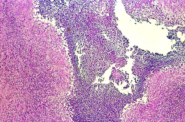
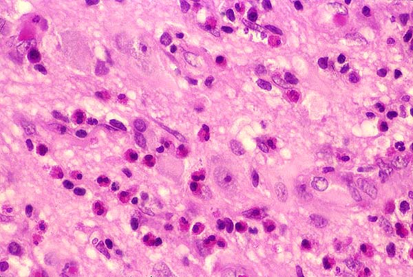
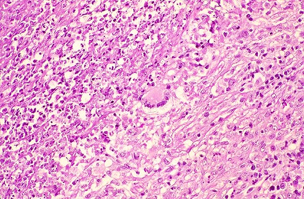
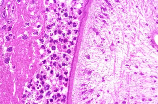
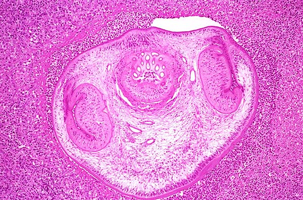
A smear preparation showed acute and chronic inflammatory cells. Histologic sections revealed necrotizing inflammatory lesions which were composed of a central cavity surrounded by an exuberant acute and chronic inflammatory infiltrate (Image 02 ) with numerous eosinophils (Image 03). In some areas, a granulomatous response with giant cells was present (Image 04). One of the cystic areas contained a parasite which was cut in cross section (Image 05). The parasite was well-preserved and demonstrated an eosinpohilic, PAS-positive tegument with microvilli. Beneath the tegument a row of palisading tegumental cells and underlying thin smooth muscle fibers were identified (Image 06). Three suckers were present in the plane of section, one of which contained hooklets, representing the rostellum (Images 07 amd 08). The surrounding brain parenchyma was gliotic. Histologic sections of the dura revealed acute and chronic inflammation with fibrosis and no identifiable parasite fragments.