MICROSCOPIC DESCRIPTION:
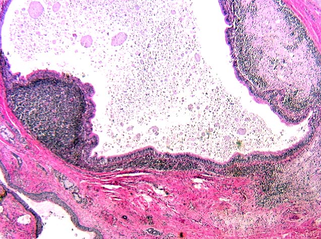
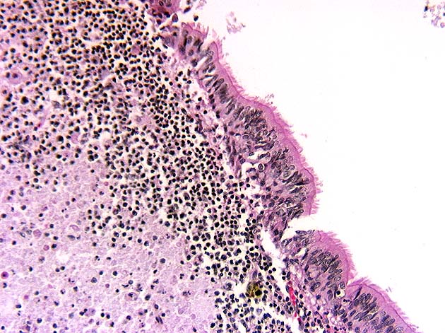
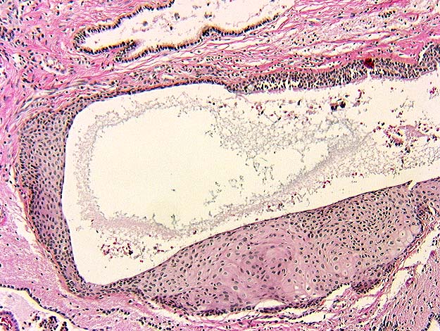
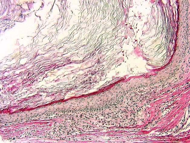
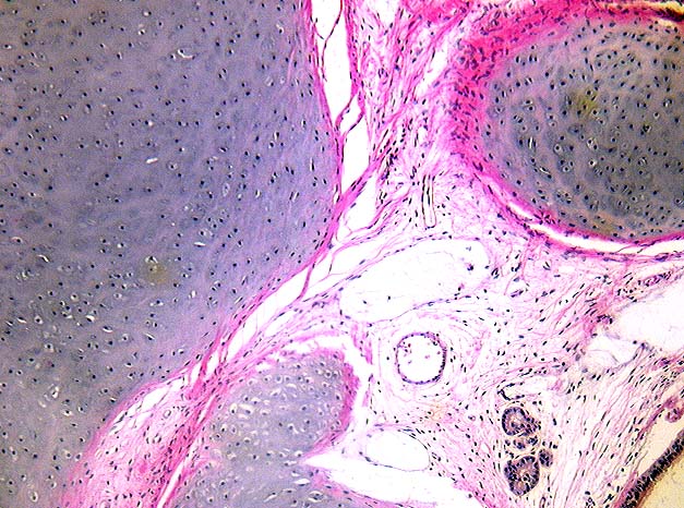
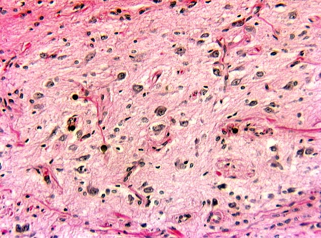
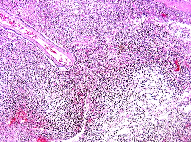
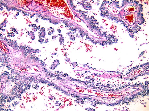
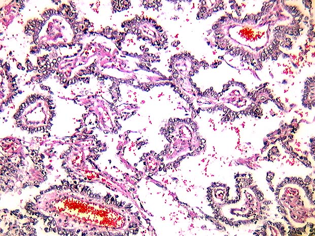
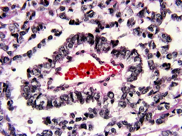
The tumor in the liver was composed of cysts with varied epithelial linings, including ciliated respiratory epithelium, (Images 07, 08) simple squamous epithelium (Image 09), keratinizing stratified squamous epithelium (Image 10), and bone were identified. Mature fibroadipose tissue and neural tissue (Image 11) and bone were identified. Mature fibroadipose tissue and neural tissue (Image 12) were present. Foci of pigmented retinal epithelium were seen. The tumor extending through the diaphragm into the lung showed a reticulated pattern (Image 13) of low cuboidal cells with some papillae intermixed (Images 14, 15). In cross section, these papillae with fibrovascular cores were lined by cuboidal cells forming Schiller-Duval bodies (Image 16). The cuboidal cells had large pleomorphic nuclei with prominent nucleoli. Some had eosinophilic hyaline globules in their cytoplasm. Similar foci of tumor were identified within the liver mass.
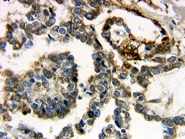
Immunoperoxidase staining for alpha fetoprotein showed cytoplasmic staining in several foci of the tumor extending through the diaphragm into the lung (Image 17).