

MICROSCOPIC DESCRIPTION:
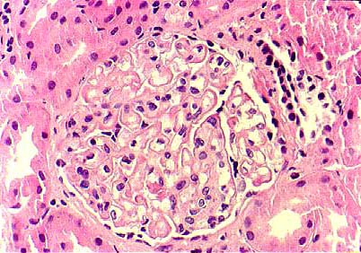
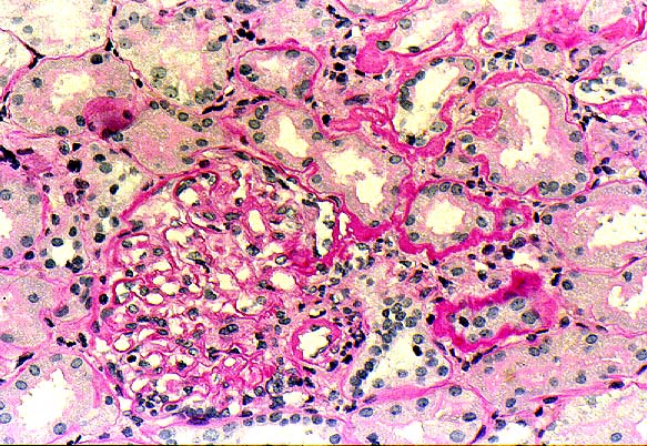
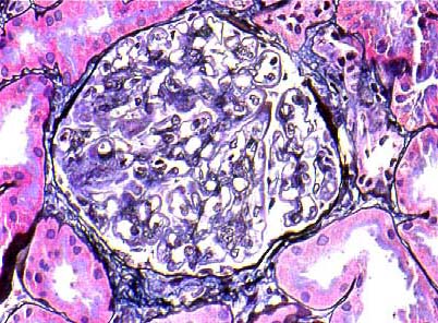
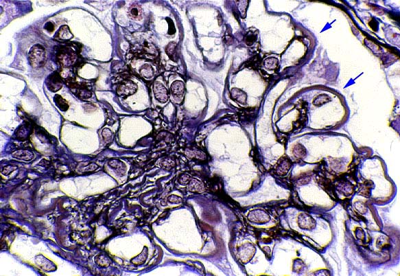
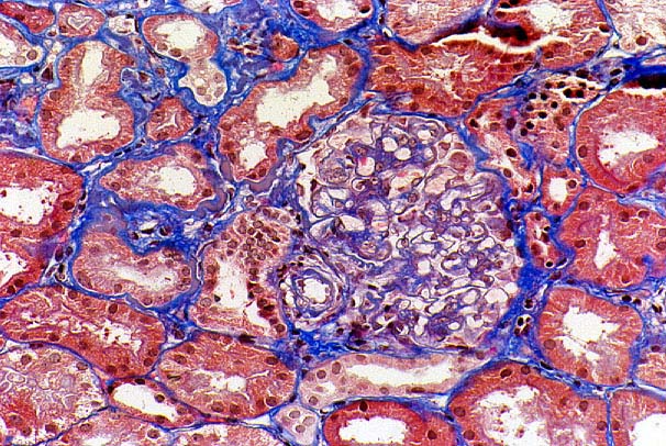
The renal biopsy tissue examined by light microscopy consisted entirely of renal cortex. The profiles of up to 36 glomeruli were identified in the paraffin, frozen and plastic sections of which two (6%) were globally sclerotic. The non-sclerotic glomeruli were largely normocellular (IMAGE 01) and exhibited diffuse, non-uniform, PAS positive (IMAGE 02) glomerular basement membrane (GBM) thickening. Methenamine silver stain showed that the non-uniform GBM thickening was due to the presence of brown semi-argyrophilic deposits bordered on each side by a thin, argyrophilic GBM producing a double contour (tram tracking--IMAGES 3A and 3B). Trichrome stain demonstrated the intramembranous deposits to be fuschinophilic (IMAGES 04A and 04B) while toluidine blue stained the deposits intermittently dark blue (IMAGE 05). No spikes nor basement membrane breaks were seen. The podocytes were slightly hypertrophied. In addition, several of the glomeruli exhibited segmental and/or global collapse with glomerular basement membrane wrinkling and focal segmental proliferation. No necrotizing lesions nor crescents were identified. The tubules were back-to-back, with several of the tubules appearing dilated. No significant tubular atrophy was identified. A few of the tubular basement membranes showed irregular, PAS positive (IMAGE 02), fuschinophilic thickening. The interstitium was unremarkable, except for a sparse, patchy lymphocytic inflammatory infiltrate. One small intrarenal artery exhibited focal mild fibroelastic intimal thickening. Other vessels appeared normal.
IMMUNOFLUORESCENCE EXAMINATION
ELECTRON MICROSCOPIC EXAMINATION