

MICROSCOPIC DESCRIPTION:
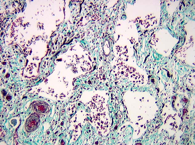
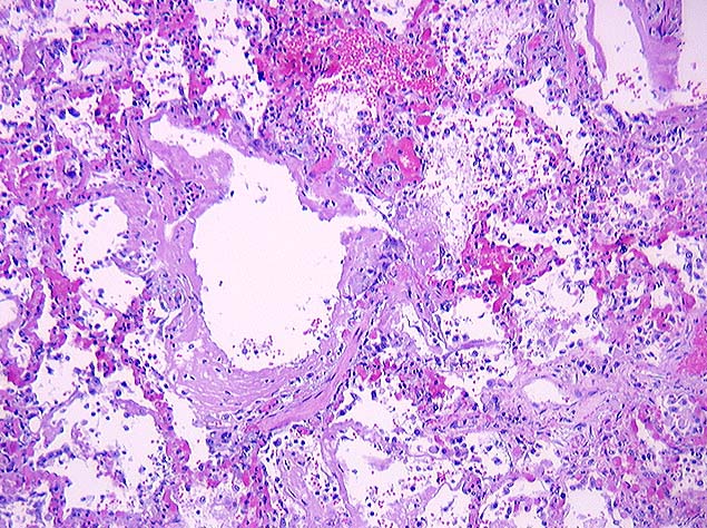
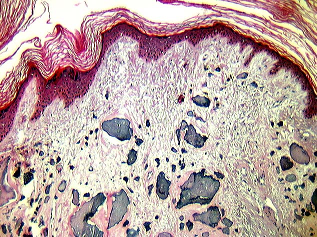
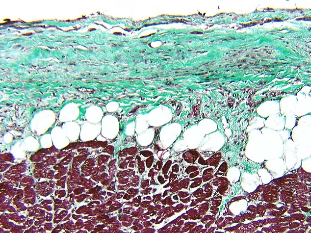
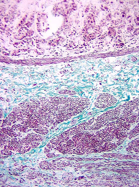
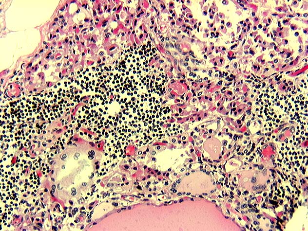
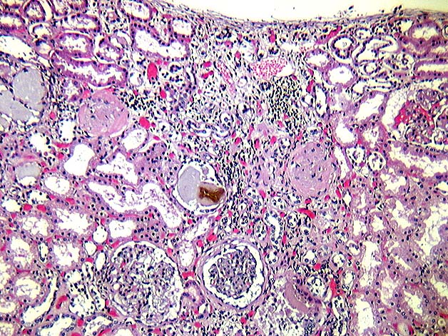
The microscopic examination revealed extensive pulmonary interstitial fibrosis (Image 04; the trichrome study revealed alveolar septal and perivascular collagen fiber deposits) with moderate to severe congestion and alveolar hemorrhage, and hypertensive changes of small arteries (Grade I hypertensive changes - Image 04). There was diffuse alveolar damage with hyaline membrane formation (Image 05) in the middle and upper lobes of the right lung. Upper lobes in both lungs showed mild emphysematous changes. There were widespread cutaneous (Image 06) and soft tissue fibrosis and calcinosis, epicardial fibrosis (Image 07, the trichrome study shows green staining of collagen fiber deposits in the epicardium), mild submucosal and smooth muscle fibrosis of GI tract (Image 08; collagen in the submucosal and intermuscular areas), fibrosis of wall of trachea (Image 09), and lymphocytic thyroiditis (Image 10). The psoas, calf, and chest muscles revealed myositis associated with multifocal muscular necrosis and atrophy, fatty and fibrous replacement (Image 11), and dystrophic calcification. Both kidneys showed scattered glomerulosclerosis and focal medial thickening of small vessels (Image 12) .