

MICROSCOPIC DESCRIPTION:
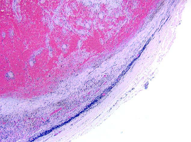
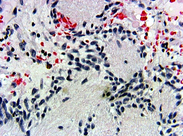
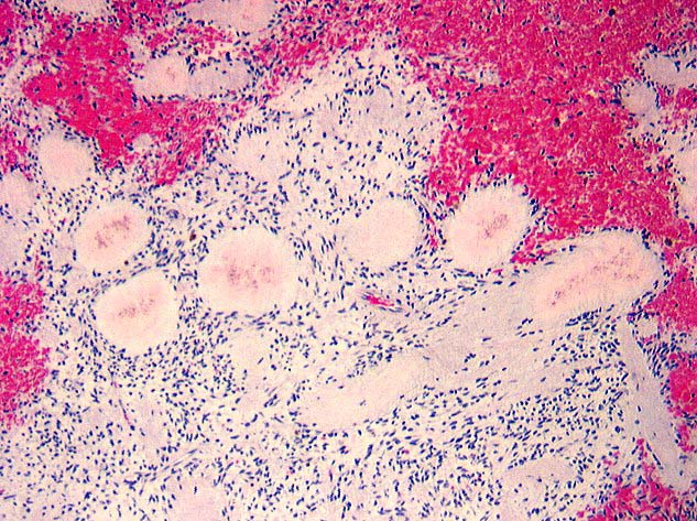
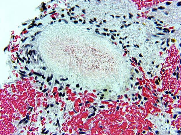
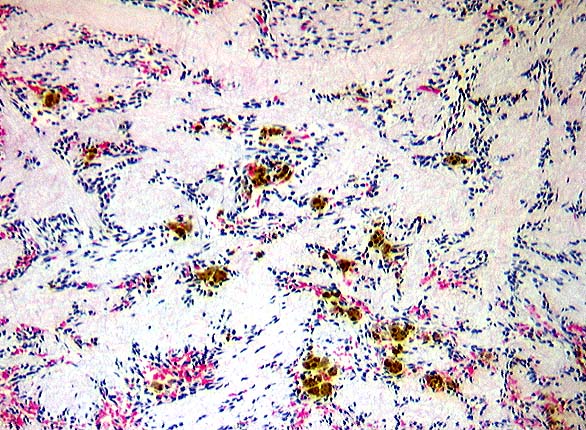
The neoplasm was composed of short interlacing fascicles of cells with uniform elongated, tapered nuclei, and fine chromatin. No mitotic activity was seen. In some areas, spindle cells with palisaded nuclei surrounded central stellate fibrillar to densely sclerotic areas. In other areas, round to stellate, deeply eosinophilic collagen-rich acellular zones (Image A, Image B and Image C) were surrounded by weakly eosinophilic rims. There was prominent hemorrhage adjacent to and within the neoplasm. A small amount of compressed normal lymphoid tissue was present at the periphery of the lesion, suggesting an intranodal origin.
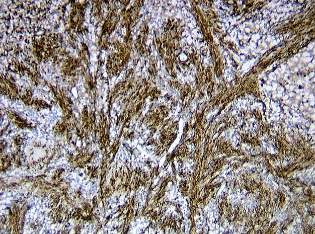
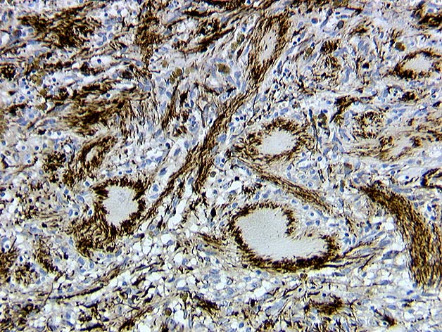
Spindle cells were immunoreactive for muscle specific actin (Image D and Image E), but not for S100, cytokeratins (AE1/AE3), Ulex europaeus, HMB45, or CD34. Collagen-rich acellular "amianthoid fibers" showed a distinctive circular pattern of actin immunoreactivity.