
MICROSCOPIC DESCRIPTION:
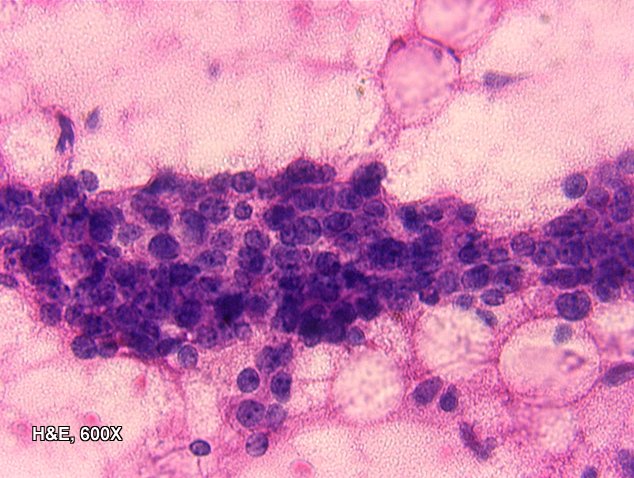
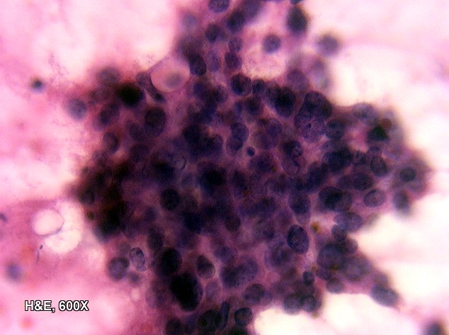
Touch preparations revealed numerous highly cellular clumps of pleomorphic spindled cells intermixed with highly cellular clumps of pleomorphic ductal cells with increased nuclear to cytoplasmic ratio, prominent nucleoli, and karyorrhectic debris.
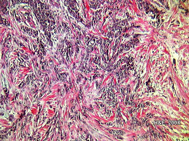
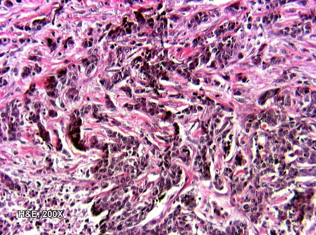

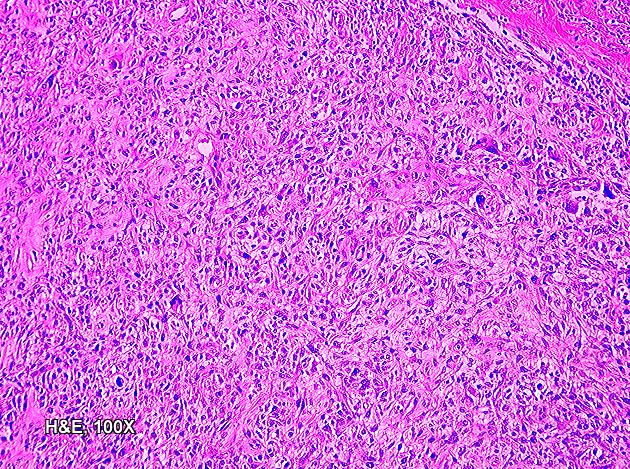
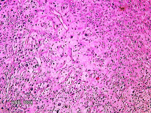
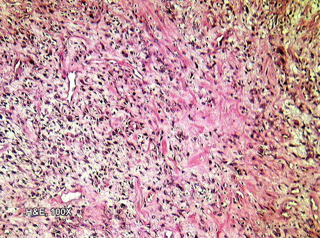
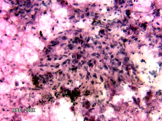
The mass was composed of areas with either glands or sheets of ductal cells (a carcinomatous component) which showed a direct transition to a spindle cell component. The glands and sheets of ductal cells showed increased nuclear to cytoplasmic ratios with pleomorphic nuclei having multiple prominent nucleoli. A high mitotic rate and foci of necrosis were seen in the carcinomatous portions. Directly adjacent to these areas of ductal carcinoma, there were areas of spindled cells with plump vesicular nuclei. Some of these areas were highly cellular with more stellate cells surrounded by amphophilic to basophilic matrix substance, which in some areas appeared chondroid. These areas also showed a high mitotic rate with foci of necrosis. Numerous multinucleated tumor giant cells were seen scattered throughout the predominantly spindled cell areas.
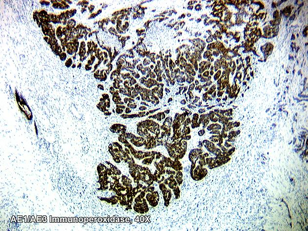
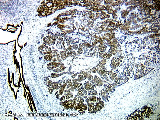
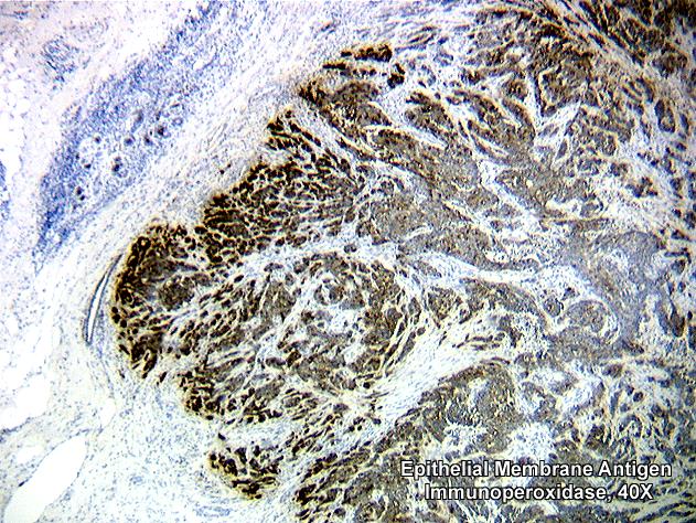


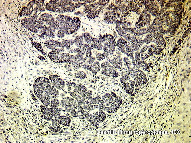
Immunoperoxidase staining of the epithelial component was positive for both cytokeratin (AE1/AE3, Cam 5.2) and epithelial membrane antigen. Vimentin and actin immunoperoxidase stains were positive in the stromal component. Desmin immunoperoxidase was negative.